Review
. 2024 May 1;134(9):e178296.
doi: 10.1172/JCI178296.

Complement regulation in the eye: implications for age-related macular degeneration

Affiliations
Review

Complement regulation in the eye: implications for age-related macular degeneration

Georgia A Wilke et al. J Clin Invest. .

Abstract

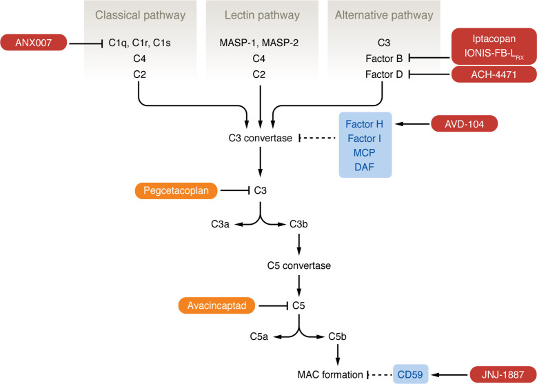
Careful regulation of the complement system is critical for enabling complement proteins to titrate immune defense while also preventing collateral tissue damage from poorly controlled inflammation. In the eye, this balance between complement activity and inhibition is crucial, as a low level of basal complement activity is necessary to support ocular immune privilege, a prerequisite for maintaining vision. Dysregulated complement activation contributes to parainflammation, a low level of inflammation triggered by cellular damage that functions to reestablish homeostasis, or outright inflammation that disrupts the visual axis. Complement dysregulation has been implicated in many ocular diseases, including glaucoma, diabetic retinopathy, and age-related macular degeneration (AMD). In the last two decades, complement activity has been the focus of intense investigation in AMD pathogenesis, leading to the development of novel therapeutics for the treatment of atrophic AMD. This Review outlines recent advances and challenges, highlighting therapeutic approaches that have advanced to clinical trials, as well as providing a general overview of the complement system in the posterior segment of the eye and selected ocular diseases.


Conflict of interest statement

Conflict of interest: RSA is a cofounder of Metro Biotech and Mobius Scientific. RSA is an advisor to Delavie Sciences, which is a subsidiary of EdenRoc Sciences, Roche, New Amsterdam Pharma, and QBioMed. RSA’s employer Washington University has intellectual property filings and patents issued that have RSA listed as an inventor.

Figures

Figure 1
Figure 1. Pathways of the complement system.
The complement system is composed of three pathways (classical, lectin, and alternative) that converge on the formation of a C3 convertase complex that is unique to each pathway. The classical pathway begins with binding of the C1 complex (composed of C1q, C1r, and C1s) to an antigen-antibody complex of pathogen surface directly; this leads to cleavage of C4 and then C2 to form the C3 convertase of the classical pathway. The lectin pathway is similar in that it begins with MBL recognizing mannose residues on a pathogen surface; this activates the MBL-associated serine proteases MASP-1 and MASP-2, which cleave C4 and C2. The alternative pathway is initiated by spontaneous hydrolysis of C3, which binds FB, leading to cleavage of FB by FD; this complex is stabilized by properdin. C3 convertase activity leads to the formation of the C5 convertase and eventually the MAC, triggering membrane destabilization of foreign material. Host complement inhibitors (light blue: FH, FI, MCP, DAF, CD59) target C3 convertase and MAC formation. FDA-approved complement inhibitors for GA (orange) are pegcetacoplan, which targets C3, and avacincaptad, which targets C5. Investigational therapies for GA (dark red) target components of the classical and alternative pathways.
Figure 2
Figure 2. The retina consists of specialized cell types organized into layers.
(A) The outer retina consists of photoreceptors, while the inner retina contains bipolar, amacrine, horizontal, Müller, and retinal ganglion cells. Bipolar cells synapse with photoreceptors and transmit their signal to ganglion cells. Horizontal and amacrine cells regulate photoreceptor and bipolar cells, respectively. Müller cells are the glial/support cells of the retina. The retina is supported by the retinal pigment epithelium (RPE). The basal lamina of the RPE forms part of Bruch’s membrane (BM), a multilayered ECM. (B) Pathological changes in early AMD occur in BM and the RPE. Basal laminar deposits appear between the RPE and the RPE basal lamina; basal linear deposits form in the inner collagenous zone of BM. Drusen are deposits beneath the RPE basal lamina; they contain cellular debris, including lipids, proteins, and complement components, such as C3, C5, and MAC (denoted by the asterisk) (74). Subretinal drusenoid deposits form anterior to the RPE and are associated with an accelerated neurodegenerative phenotype in AMD.
Figure 3
Figure 3. The retina in AMD.
(A) In neovascular AMD, it is hypothesized that choriocapillaris atrophy leads to ischemia of the RPE, which triggers VEGF secretion and the growth of abnormal choroidal blood vessels. These vessels breach BM and grow in the sub-RPE or subretinal space, causing accumulation of subretinal and intraretinal fluid. (B) In atrophic AMD, it is thought that some primary insult leads to RPE degeneration, which causes choriocapillaris atrophy due to the role of the RPE in supporting choriocapillaris function. As the RPE degenerates, the overlying photoreceptors die. In both types of AMD, there is choriocapillaris atrophy and RPE degeneration, though the sequence of events in each disease may be different. In terms of complement activity in AMD, increased concentrations of C3, C3a, Bb, FB, and FD have been detected within BM and choriocapillaris of human donor eyes with AMD (denoted by asterisks) (100). Cadaver studies have found MAC deposition in the RPE and choriocapillaris of patients with the Y402H polymorphism in CFH regardless of whether AMD changes are present (88, 89). The Y402H polymorphism is believed to contribute to AMD pathogenesis primarily through its effect on FHL-1, as FHL-1 is the major complement regulator of BM (83, 84).

References

    1. Medawar PB. Immunity to homologous grafted skin; the fate of skin homografts transplanted to the brain, to subcutaneous tissue, and to the anterior chamber of the eye. Br J Exp Pathol. 1948;29(1):58–69. - PMC - PubMed
    1. Luke Qi Jiang, Streilein JW. Immune privilege extended to allogeneic tumor cells in the vitreous cavity. Invest Ophthalmol Vis Sci. 1991;32(1):224–228. - PubMed
    1. Benhar I, et al. The privileged immunity of immune privileged organs: the case of the eye. Front Immunol. 2012;3:296. doi: 10.3389/fimmu.2012.00296. - DOI - PMC - PubMed
    1. Sohn JH, et al. Tolerance is dependent on complement C3 fragment iC3b binding to antigen-presenting cells. Nat Med. 2003;9(2):206–212. doi: 10.1038/nm814. - DOI - PMC - PubMed
    1. Perez VL, Caspi RR. Immune mechanisms in inflammatory and degenerative eye disease. Trends Immunol. 2015;36(6):354–363. doi: 10.1016/j.it.2015.04.003. - DOI - PMC - PubMed

Publication types

Substances