Review
. 2024 Jun 13;37(2):e0003423.
doi: 10.1128/cmr.00034-23. Epub 2024 May 1.

An updated list of eumycetoma causative agents and their differences in grain formation and treatment response

Affiliations
Review

An updated list of eumycetoma causative agents and their differences in grain formation and treatment response

Wendy W J van de Sande et al. Clin Microbiol Rev. .

Abstract

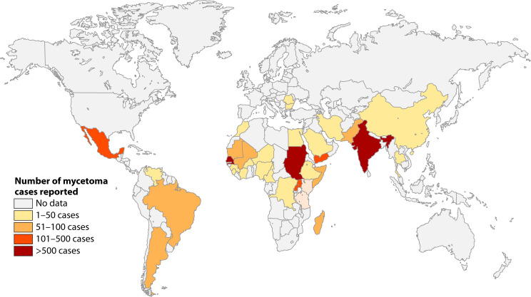
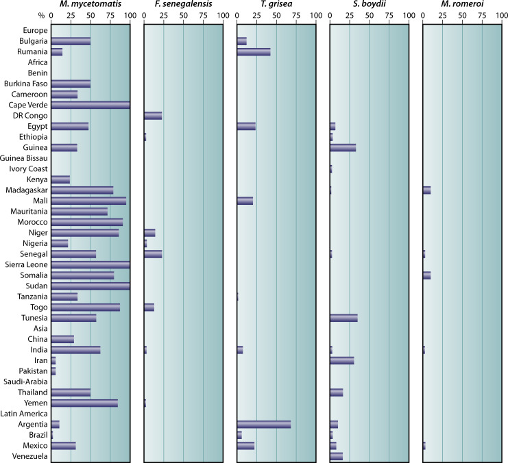
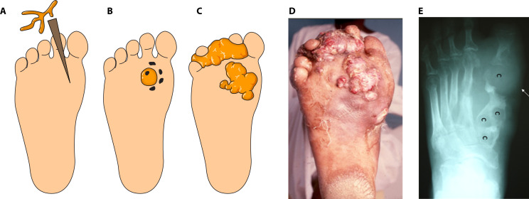
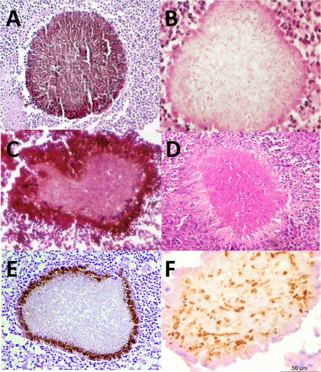
SUMMARYIn 2023, the World Health Organization designated eumycetoma causative agents as high-priority pathogens on its list of fungal priority pathogens. Despite this recognition, a comprehensive understanding of these causative agents is lacking, and potential variations in clinical manifestations or therapeutic responses remain unclear. In this review, 12,379 eumycetoma cases were reviewed. In total, 69 different fungal species were identified as causative agents. However, some were only identified once, and there was no supporting evidence that they were indeed present in the grain. Madurella mycetomatis was by far the most commonly reported fungal causative agent. In most studies, identification of the fungus at the species level was based on culture or histology, which was prone to misidentifications. The newly used molecular identification tools identified new causative agents. Clinically, no differences were reported in the appearance of the lesion, but variations in mycetoma grain formation and antifungal susceptibility were observed. Although attempts were made to explore the differences in clinical outcomes based on antifungal susceptibility, the lack of large clinical trials and the inclusion of surgery as standard treatment posed challenges in drawing definitive conclusions. Limited case series suggested that eumycetoma cases caused by Fusarium species were less responsive to treatment than those caused by Madurella mycetomatis. However, further research is imperative for a comprehensive understanding.

Keywords: biofilm; diagnosis; grain; itraconazole; molecular diagnostics; mycetoma; neglected tropical disease; susceptibility.


Conflict of interest statement

The authors declare no conflict of interest.

Figures

Fig 1
Fig 1
Number of eumycetoma cases reported based on the metadata used for publications (3, 5) using references (6–94).
Fig 2
Fig 2
Distribution of the most common causative eumycetoma agents per country. For each country, the percentage of M. mycetomatis, F. senegalensis, T. grisea, S. boydii, and M. romeroi were calculated by the following formula (number of cases per selected species/total number of eumycetoma cases reported in that country × 100) and displayed in the corresponding panels.
Fig 3
Fig 3
Eumycetoma development. (A) The fungal causative agent is introduced into the subcutaneous tissue via a minor trauma such as a thorn prick. (B) Inside the tissue, the fungus will form a grain. (C) Subcutaneous nodules will form, which will further extend into the subcutaneous tissue. (D) Picture of a mycetoma lesion in real life. (E) The eumycetoma causative agents will invade the bone and cavities (c) will form.
Fig 4
Fig 4
Histology of grains. (A) M. mycetomatis; (B) S. boydii picture reprinted from reference (201) with permission of the publisher (McGraw Hill); (C) F. senegalensis, picture reprinted from reference (202); (D) Fusarium, picture reprinted from reference (203) with permission of the publisher (copyright 2016 Blackwell Verlag GmbH); (E) T. grisea, picture reprinted from reference (204) with permission; (F) A. flavus, stained with antibodies against Aspergillus, picture reprinted from reference (107) with permission of the publisher (copyright 2015 Blackwell Verlag GmbH).
Fig 5
Fig 5
Grain formation over time. (A) Fungal cells are recognized by the host via pathogen receptor proteins. (B) Hemocytes will then agglutinate around the fungal hyphae, and host cells will be cross-linked to each other and attach to the fungal cells. The fungal cells will excrete zincophores and siderophores to obtain nutrients. (C) Degranulation of the host cells will occur, and reactive oxygen species will be produced. In order to protect itself, the fungus will produce melanin and trehalose. The host will form a capsule surrounding the granuloma. (D) In the final stage, the fungus will completely disintegrate the host cells and form the cement material. The extracellular matrix will be melanized. Although the steps in grain formation are similar between M. mycetomatis and F. senegalensis, the timing is not. (E) A M. mycetomatis grain in G. mellonella 4 hours after infection. Cement material is forming. (F) A M. mycetomatis grain in G. mellonella 24 hours after infection. Cement material is formed, and only a few host cells are still found within the grain. (G) A mature M. mycetomatis grain in G. mellonella larvae after 3 days. At this time no host cells are present in the cement material. The cement material is melanized, and a capsule surrounds the grain. (H) A massive influx of immune cells toward the M. mycetomatis grains 7 days after infection. (I) No grain is present in G. mellonella larvae infected with F. senegalensis 4 hours after infection. Only loose hyphae are noted. (J) At 24 hours after infection, the first signs of grain formation are noted. There is no cement material yet, and melanization is not noted. (K) F. senegalensis grain at 3 days after infection, cement material is forming, and some melanization of the grain is noted. Also, a capsule is forming. (L) At 7 days after infection, a mature F. senegalensis grain in G. mellonella is noted. Panels A–D are based on reference (194), panels E–H are reprinted from reference (174) (published under a Creative Commons license), and panels I–L are reprinted from reference (213) (published under a Creative Commons license).
Fig 6
Fig 6
Identification of causative agents by growth characteristics. Assimilation of or growth on potassium gluconate (PG), potassium 2-keto-gluconate (P2KG), methyl-D-glucopyranoside (MDG), actidione (ACT), or L-sorbose can help in differentiating the most common causative agents of black grain mycetoma. Adapted from references (94, 220).

Similar articles

Cited by

References

    1. WHO . 2016. WHA69.21: assessing the burden of mycetoma. WHO, Geneva.
    1. Zijlstra EE, van de Sande WWJ, Welsh O, Mahgoub ES, Goodfellow M, Fahal AH. 2016. Mycetoma: a unique neglected tropical disease. Lancet Infect Dis 16:100–112. doi:10.1016/S1473-3099(15)00359-X - DOI - PubMed
    1. van de Sande W, Fahal A, Ahmed SA, Serrano JA, Bonifaz A, Zijlstra E, eumycetoma working group . 2018. Closing the mycetoma knowledge gap. Med Mycol 56:153–164. doi:10.1093/mmy/myx061 - DOI - PubMed
    1. WHO . 2023. WHO fungal priority pathogens list to guide research, development and public health action
    1. van de Sande WWJ. 2013. Global burden of human mycetoma: a systematic review and meta-analysis. PLoS Negl Trop Dis 7:e2550. doi:10.1371/journal.pntd.0002550 - DOI - PMC - PubMed

Substances

LinkOut - more resources