. 2024 Jun;234(1):33-44.
doi: 10.1007/s00427-024-00716-2. Epub 2024 May 1.

From bristle to brain: embryonic development of topographic projections from basiconic sensilla in the antennal nervous system of the locust Schistocerca gregaria

Affiliations

From bristle to brain: embryonic development of topographic projections from basiconic sensilla in the antennal nervous system of the locust Schistocerca gregaria

George Boyan et al. Dev Genes Evol. 2024 Jun.

Abstract

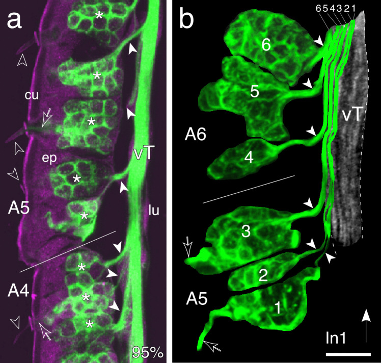
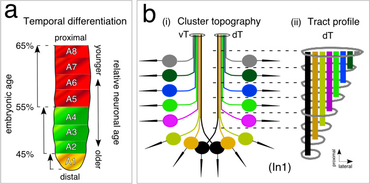
The antennal flagellum of the locust S. gregaria is an articulated structure bearing a spectrum of sensilla that responds to sensory stimuli. In this study, we focus on the basiconic-type bristles as a model for sensory system development in the antenna. At the end of embryogenesis, these bristles are found at fixed locations and then on only the most distal six articulations of the antenna. They are innervated by a dendrite from a sensory cell cluster in the underlying epithelium, with each cluster directing fused axons topographically to an antennal tract running to the brain. We employ confocal imaging and immunolabeling to (a) identify mitotically active sense organ precursors for sensory cell clusters in the most distal annuli of the early embryonic antenna; (b) observe the subsequent spatial appearance of their neuronal progeny; and (c) map the spatial and temporal organization of axon projections from such clusters into the antennal tracts. We show that early in embryogenesis, proliferative precursors are localized circumferentially within discrete epithelial domains of the flagellum. Progeny first appear distally at the antennal tip and then sequentially in a proximal direction so that sensory neuron populations are distributed in an age-dependent manner along the antenna. Autotracing reveals that axon fasciculation with a tract is also sequential and reflects the location and age of the cell cluster along the most distal annuli. Cell cluster location and bristle location are therefore represented topographically and temporally within the axon profile of the tract and its projection to the brain.

Keywords: Antenna; Basiconic sensilla; Embryogenesis; Locust; Topographic projections.


Conflict of interest statement

The authors declare no competing interests.

Figures

Fig. 1
Fig. 1
Cuticular bristles and their innervation on the antenna of a first instar (In1) locust. a Low power photomicrograph of the ventral cuticular surface of the six most distal segments or annuli (A6, A5, A4, A3, A2, A1; terminology from Chapman 2002) of the antenna. Prominent bristles (white arrowheads) are typically found near the distal border of each segment. Distal is to the bottom. b Photomicrograph at higher power shows the bristles in the regions indicated panel a (open white arrowheads) to be of the basiconic-type (see Suppl. Figure 1). According to Chapman (2002), these are only found on the six most distal annuli and have a consistent location in each. Other sensilla present in the images but not part of this study include coeloconic (open black arrowhead) and campaniform sensilla (black arrowheads) (for details of receptor morphologies, see Slifer et al. , ; Chapman and Greenwood ; Ochieng et al. ; Chapman 2002). c, d Confocal images following neuron-specific α-HRP immunolabeling (green) show clusters of sensory cells (white stars) in the epidermis (epi) associated with basiconic-type bristles on the cuticle (cu) of annuli A6 (panel c) and A4 (panel d) in longitudinally sectioned antennae from a first instar locust. Cuticular and epidermal regions autofluoresce (false color magenta). A fasciculated bundle of HRP-positive neurites (open/white arrow) projects from each epidermal cell cluster and terminates as a dendrite (white arrowhead) at the tip within each bristle, consistent with the innervation of basiconic-type sensilla (see Suppl. Figure 1). Fasciculated axons (open white arrowhead) exit each cell cluster en route to the antennal nerve (not imaged here but see Fig. 6). Scale bar represents 180 µm in a, 45 µm in b, 15 µm in c, d
Fig. 2
Fig. 2
Ontogeny and development of sensory cell lineages in apical segments of the embryonic antenna. a Culturing with the S-phase label EdU (white) and co-labeling with the nuclear stain DAPI (blue) reveals differentiating SOPs in the epithelium (ep) of annuli A1, A2, A3, and A4 at 30% of embryogenesis. Unidentified differentiating cells are also present in the lumen (lu) of A1 and A4. Distal is to the bottom. b Triple labeling against Lachesin (α-Lach, red), against the S-phase label EdU (white), and using the nuclear stain DAPI (blue) shows an EdU-labeled, Lach-positive SOP (white arrowhead) differentiating at the tip of a column of epithelial (ep) cells in A1 of repeat preparations (i, ii) at 31% of embryogenesis. c Confocal image following double labeling against Lachesin (α-Lach, red) and the proliferative cell marker phospho-histone 3 (α-PH3, white) at 33% of embryogenesis reveals mitotically active precursor cells restricted to the Lach-positive epithelial domains of A1, A2, A3, and A4. d Confocal images of antennal annuli A4 (i) and A2 (ii) in cross-section at 45% of embryogenesis following double labeling against Lachesin (α-Lach, red) and anti-phospho-histone 3 (α-PH3, white). PH3-positive mitotically active precursors are located circumferentially in the epithelial domain (ep) of each annulus, as are Lach-positive differentiating cell clusters (open white arrowheads). A single mitotically active cell is located in the lumen (lu) of A2. Panel A4 modified from Boyan and Ehrhardt (2020). e Confocal images following triple labeling against Lachesin (α-Lach, red), horseradish peroxidase (α-HRP, green), and phospho-histone 3 (α-PH3, white) reveal SOPs and associated HRP-positive/Lach-positive cells in annulus A2 of the embryonic antenna. At 38% (i), a representative Lach-positive proliferative SOP (white star) is associated with three Lach-/HRP-positive cells (white crosses). The SOP is typically linked to the cuticle (cu, dashed white line) by an epithelial foot (white arrowhead; terminology follows Locke and Huie 1981). Modified from Boyan and Ehrhardt (2020). At 48% (ii), a mitotically active SOP (white star) has detached from the epithelial cell layer and is associated with at least six Lach-/HRP-positive cells (white crosses). f Confocal image following double labeling against horseradish peroxidase (α-HRP, green) and phospho-histone 3 (α-PH3, white) at 55% of embryogenesis reveals mitotically active SOPs (white stars) each associated with a cluster of HRP-positive cells (white crosses) in annulus A4 of the antenna. Axonal processes (open white arrowheads) project centrally onto the ventral tract (vT), one of the two axon tracts projecting proximally to the antennal base. Fused dendritic processes (white arrowheads) from HRP-positive cell clusters associated with SOPs (located behind the more peripheral SOP) project peripherally towards the cuticular edge of the antenna (cu, dashed white) where they may become associated with sensilla that develop later (see Suppl. Figure 2). Scale bar represents 25 µm in a, 18 µm in b, 25 µm in c, 60 µm in d, 20 µm in e, 30 µm in f
Fig. 3
Fig. 3
Immunolabeling against horseradish peroxidase (α-HRP) shows that sensory cell clusters appear in a distal to proximal direction along the antenna during embryogenesis. The arrow points to the antennal base. a At 45%, fluorescence photomicrograph shows sensory cell clusters (white arrowheads) in A1 of the flagellum (Fl), with a single cluster present unilaterally in A4. Neurons from these clusters project axons topographically onto either the ventral (vT) or dorsal (dT) antennal tracts running proximally to the antennal base. Cell clusters of Johnston’s organ (Jo) are also present in the pedicellum (Pe). b At 53%, fluorescence photomicrograph shows increasing numbers of cell clusters in A1, A2, and A4, while isolated sensory cell clusters are now evident in A6 and A8 (white arrowheads) along with their peripherally projecting fused dendrites. c At 56%, the confocal image reveals greater numbers of cell clusters are present throughout A1, A2, and A4 and also appear in A3. Note that cell cluster numbers in more proximal annuli A5–A8 are essentially similar to those of 53%. d At 65%, the confocal image shows cell cluster numbers have increased basally and now fill the epithelia of A1–A8. Panels a and b modified from Boyan and Williams (2004). Scale bar represents 90 µm in a, 165 µm in bd
Fig. 4
Fig. 4
Developing patterns of fasciculation and topological organization of projections from sensory cell clusters to the two primary axon tracts that project proximally to the antennal base. Confocal images follow labeling against horseradish peroxidase (α-HRP). Ventral cell clusters and axon tracts are shown in false color magenta and dorsal cell clusters and tracts in false color cyan. Distal is to the bottom in all panels. a At 43% of embryogenesis, cell clusters (white stars) have appeared in distal segments A1, A2, and A4 of the antenna. Clusters in A1 have generated axons (white arrowheads) that project topologically onto the primary tract system (vT, dT) of the antenna. b At 48%, sensory cell clusters (white stars) arrayed in the epithelium (ep) are clearly associated with either a ventral (magenta) or dorsal (cyan) axon tract. Each tract runs along the border between the lumen (lu) and epithelium towards the antennal base. Dendrites (open white arrows) from some cell clusters can be seen to extend towards the cuticle (dashed white) but bristles are not yet evident. c Confocal image montaged from optical stacks collated according to depth demonstrates that at 85% of embryogenesis distal HRP-positive cell clusters (white stars label some) located ventrally (magenta) or dorsally (cyan) in the epithelium (ep) of A1 and A2 project axons (white arrowheads) topologically onto respective ventral (vT, magenta) or dorsal (dT, cyan) antennal tracts. Note the symmetrical projection patterns. Fused HRP-positive dendrites from these clusters innervate ventral (magenta) or dorsal (cyan) cuticular bristles (open white arrowheads). Scale bar represents 33 µm in a, 35 µm in b, 25 µm in c
Fig. 5
Fig. 5
Axons from clusters of cells associated with basiconic bristles project topographically into a tract of the antennal nervous system. a A confocal image of a longitudinal section through the antenna in side view following immunolabeling with anti-horseradish peroxidase (α-HRP, green) at 95% of embryogenesis shows an array of sensory cell clusters (white stars) in the epithelium (ep) of segments A4 and A5 of the flagellum (white line indicates approximate border). Cuticular/epithelial autofluorescence appears magenta. Sensory dendrites (open white arrows) are seen innervating basiconic bristles (open/white arrowheads) on the cuticle (cu). Bundled axons (white arrowheads) from each cell cluster exit the epithelium and join the ventral nerve tract (vT) in the lumen (lu) in sequential order from distal to proximal (direction indicated by the white arrow in panel b). b Higher power confocal image of a longitudinal section through the antenna (side view) of a first instar (In1) locust following labeling against horseradish peroxidase (α-HRP, green) shows computer-based autotracings of six HRP-positive sensory cell clusters numbered sequentially from distal to proximal in the epithelium of A5 and A6 (white line is the approximate border). Autotraced axons from each cluster (white arrowheads) join the α-HRP-labeled vT (false color gray, outlined dashed white) topographically according to the location of the cell cluster in the epithelium (axon and cell cluster numbers correspond). Axons from distal clusters lie medially in the tract, and those from progressively more proximal clusters add laterally to the tract in sequence producing a topographic effect akin to tree rings. Dendrites (open white arrows) from cell clusters 1 and 3 innervating basiconic bristles in A5 are also imaged. Scale bar represents 20 µm in a, 10 µm in b
Fig. 6
Fig. 6
Schematics (not to scale) summarize the spatial and temporal organization of sensory cell clusters and their axonal projections in the developing antenna. a Temporal differentiation. Cell clusters appear in the epithelium in a distal to proximal direction providing a temporal topology to cluster organization. Cell clusters first appear in annulus A1 (yellow), then in A2–A4 (green), and finally in A5–A8. Patterns are complete in these annuli at 65% of embryogenesis. The pattern suggests that neurons from distal cell clusters are older than those in more proximal annuli. b (i) Cluster topography. Cell clusters (colored circles) receiving sensory input from cuticular basiconic-type bristles (black arrows) are arrayed in the epithelium of the distal antenna. Note that the depicted cluster location is representative but in sum covers the most distal six annuli. Axons from a given cluster project topographically to either a ventral (vT) or dorsal (dT) tract and join the tract commensurate with the location of the cluster along the array. (ii) Tract profile. A longitudinal sketch of the dorsal tract (dT, also applies to the vT) taken at different levels along the antenna summarizes axon topology in the tract. Clusters and axons are color matched. The tract increases in diameter as younger axons from progressively more proximal clusters join. The location of axons in the tract is stereotypic and so reflects the topography and temporal topology of cell clusters in the epithelium

Similar articles

References

    1. Adams RR, Maiato H, Earnshaw W, Carmena M. Essential roles of Drosophila inner centromere protein (INCENP) and aurora B in histone H3 phosphorylation, metaphase chromosome alignment, kinetochore disjunction, and chromosome Segregation. J Cell Biol. 2001;153:865–879. doi: 10.1083/jcb.153.4.865. - DOI - PMC - PubMed
    1. Ai H, Nishino H, Itoh T. Topographic organization of sensory afferents of Johnston’s organ in the honeybee brain. J Comp Neurol. 2007;502:1030–1046. doi: 10.1002/cne.21341. - DOI - PubMed
    1. Bentley D, Keshishian H, Shankland M, Torian-Raymond A. Quantitative staging of embryonic development of the grasshopper, Schistocerca nitens. J Embryol Exp Morphol. 1979;54:47–74. - PubMed
    1. Boronat-Garcia A, Iben J, Dominguez-Martin E, Stopfer M. Identification and analysis of odorant receptors expressed in the two main olfactory organs, antennae and palps, of Schistocerca americana. Sci Rep. 2022;12:22628. doi: 10.1038/s41598-022-27199-3. - DOI - PMC - PubMed
    1. Boyan G, Ehrhardt E. Ontogeny of pioneer neurons in the antennal nervous system of the grasshopper Schistocerca gregaria. Dev Genes Evol. 2017;227:11–23. doi: 10.1007/s00427-016-0565-0. - DOI - PubMed

LinkOut - more resources