

A case of dumbbell-shaped accessory scrotum with concomitant lipoma
- PMID: 38691310
- PMCID: PMC11063015
- DOI: 10.1186/s40792-024-01906-w
A case of dumbbell-shaped accessory scrotum with concomitant lipoma
Abstract
Background: Accessory scrotum is a congenital scrotal anomaly that is usually located anterior to the anus and frequently presents with a lipoma in a bead-like shape. Herein, we present an unusual case of an accessory scrotum with a lipoma connected by a narrow stalk and located posterior to the anus.
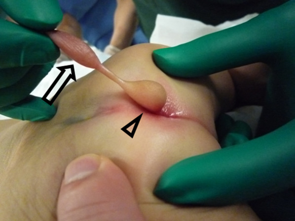
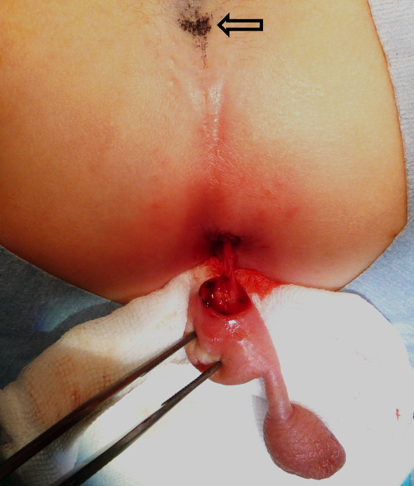
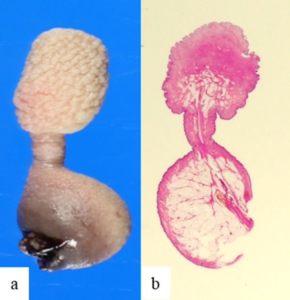
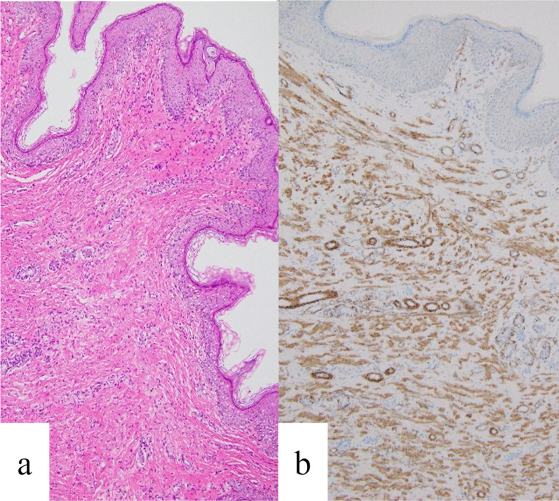
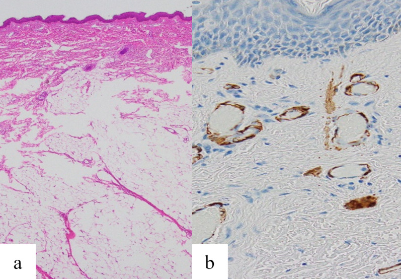
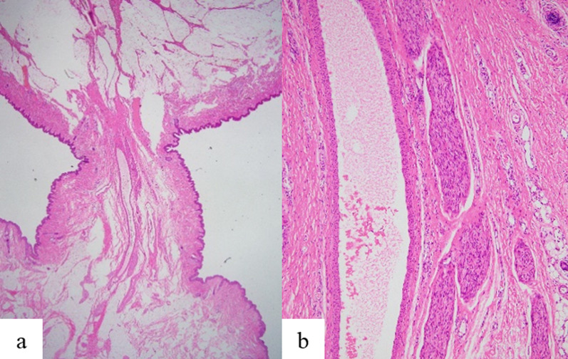
Case presentation: A 1-month-old boy was referred to our hospital for a perineal mass present at birth. He was born at 37 weeks and 2 days, with a birth weight of 2962 g. No abnormalities occurred during the perinatal period, and the birth was uneventful. The mass had an unusual shape, comprising two masses connected by a narrow stalk. The base of the mass was posterior to the anus and was connected to the rectal mucosa. The proximal mass was elastic and soft without skinfolds, whereas the distal mass was elastic and soft with a scrotum-like skinfolds. Magnetic resonance imaging showed no spina bifida. High-intensity adipose tissues in both masses and low-intensity vessels or fibrous stroma in cord-like structures between the two masses were found on T2-weighted images. At 3 months of age, the patient underwent resection in the prone jackknife position. No tumorous lesions were connected to the mass on the rectal and coccyx sides, and the mass was completely removed, preserving the anal sphincter. Histologically, the distal mass had characteristics of a scrotum, whereas the proximal mass was exclusively a lipoma. The connecting stalk had normal skin structures and a blood vessel with parallel-running nerve bundles. The postoperative course was uneventful, and the patient was discharged on postoperative day 6.
Conclusions: This case of accessory scrotum was unusual in its location and the presence of a stalk connecting the accessory scrotum and lipoma. The mechanism underlying accessory scrotum development remains unclear, and our report may impact the discourse regarding the embryological development of the accessory scrotum.
Keywords: Accessory scrotum; Lipoma; Neonate; Stalk.
© 2024. The Author(s).
Conflict of interest statement
The authors have no conflicts of interest to declare.
Figures
References
-
- Ikegami M, Urao M, Tanaka N, Ashizawa K, Ogura K, Matsumoto T. Diagnosing the accessory scrotum near the anus diagnosed by desmin staining: a case report. Jpn J Pediatr Urol. 2022;31:79–83.
LinkOut - more resources
Full Text Sources
