. 2024 May 3;10(18):eadl1922.
doi: 10.1126/sciadv.adl1922. Epub 2024 May 1.

Exchange of subtelomeric regions between chromosomes 4q and 10q reverts the FSHD genotype and phenotype

Affiliations

Exchange of subtelomeric regions between chromosomes 4q and 10q reverts the FSHD genotype and phenotype

Yinxing Ma et al. Sci Adv. .

Abstract

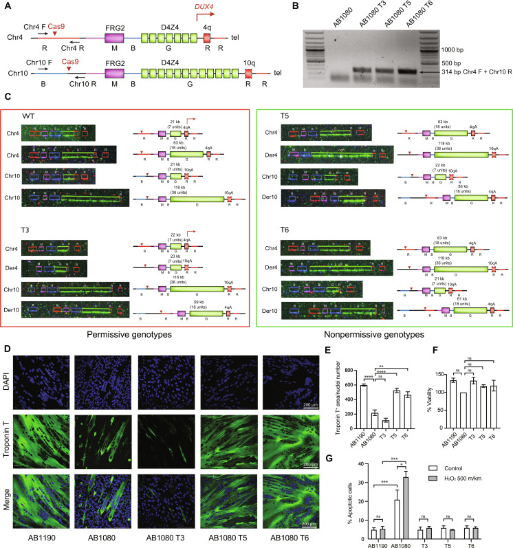
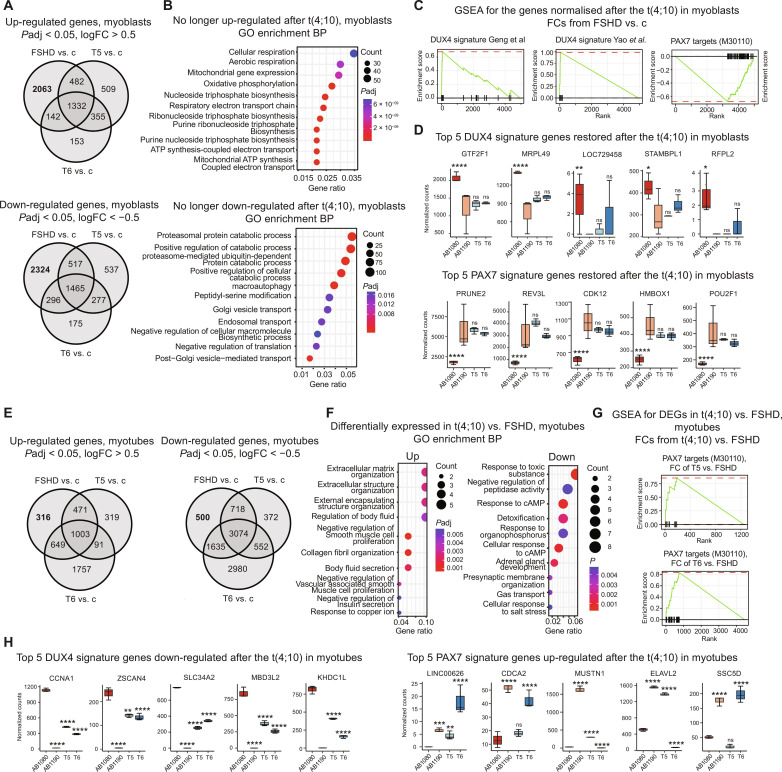
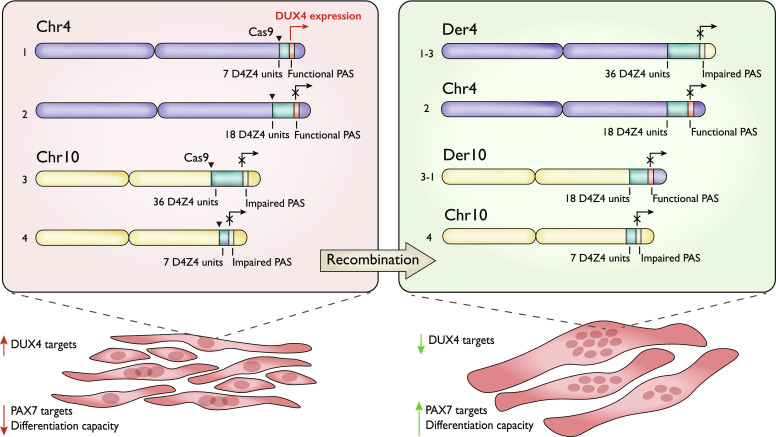
The most common form of facioscapulohumeral dystrophy (FSHD1) is caused by a partial loss of the D4Z4 macrosatellite repeat array in the subtelomeric region of chromosome 4. Patients with FSHD1 typically carry 1 to 10 D4Z4 repeats, whereas nonaffected individuals have 11 to 150 repeats. The ~150-kilobyte subtelomeric region of the chromosome 10q exhibits a ~99% sequence identity to the 4q, including the D4Z4 array. Nevertheless, contractions of the chr10 array do not cause FSHD or any known disease, as in most people D4Z4 array on chr10 is flanked by the nonfunctional polyadenylation signal, not permitting the DUX4 expression. Here, we attempted to correct the FSHD genotype by a CRISPR-Cas9-induced exchange of the chr4 and chr10 subtelomeric regions. We demonstrated that the induced t(4;10) translocation can generate recombinant genotypes translated into improved FSHD phenotype. FSHD myoblasts with the t(4;10) exhibited reduced expression of the DUX4 targets, restored PAX7 target expression, reduced sensitivity to oxidative stress, and improved differentiation capacity.


Figures

Fig. 1.
Fig. 1.. Genotype and phenotype characterization of the recombinant clones.
(A) Schematic representation of the 4q35 and 10q26 loci and the location of the primers to detect the t(4;10)(q35;q26). Chromosomal regions are colored according to the colors of the fluorescent hybridization probes from the FiberVision assay (R, red; B, blue; G, green; M, magenta). The dashed line outlining the 10qA allele represents the impaired polyadenylate signal. The red errors point to the guide RNA target sequences. (B) PCR products obtained with the Chr4 F/Chr10 R primer pair in the maternal AB1080 cells and the recombinant clones. The presence of the 314-bp hybrid chr4-chr10 product indicates successful t(4;10)(q35;q26) induction. (C) FiberVision FSHD assay characterizing the structure of the FSHD locus in the maternal AB1080 cells and the recombinant clones T3, T5, and T6. The green fluorescent probe (G) hybridizes to the D4Z4 units to determine the length of the D4Z4 array; the most proximal probe distinguishes between chr4 (red, R) and chr10 (blue, B); the most distal probe distinguishes between the A (red, R) and B (blue, B) chr4/chr10 alleles. In the AB1080 cell line, both copies of chr4 and chr10 carry the A variant. (D) Myotubes formed by control, FSHD, and the recombinant AB1080 T3, T5, and T6 myoblasts, fourth day of differentiation; staining with the monoclonal antibodies against skeletal troponin T (green) and 4′,6-diamidino-2-phenylindole (DAPI) (blue). Representative fields. (E) Differentiation efficiency assessment; troponin T. positive area normalized to the nuclei number, means ± SEM. N = 6. (F) Cell viability assessment in the control, FSHD, and the recombinant T3, T5, and T6 myoblasts; means ± SEM, N = 3. (G) Apoptosis assessment in the control, FSHD, and the recombinant T3, T5, and T6 myoblasts following the oxidative stress induction by 500 mkm of H2O2; means ± SEM, N = 3.
Fig. 2.
Fig. 2.. Transcriptome profiling of the recombinant clones.
(A) Up-regulated and down-regulated genes in FSHD, T5, or T6 myoblasts versus control myoblasts. (B) GO Biological Process enrichment for genes no longer deregulated in both translocation clones at the myoblast stage. (C) Gene Set Enrichment Analysis (GSEA) against the DUX4 signature gene sets from Geng et al. (3) and Yao et al. (2) and PAX7 targets gene set (M30110) (44) for the genes no longer deregulated in both translocation clones at the myoblast stage. The genes were thresholded by Padj < 0.05 and ranked by the log2FC from the FSHD versus control myoblasts comparison. (D) DUX4 and PAX7 signature genes restored in both translocation clones. Top 5 genes by Padj in the FSHD versus control myoblasts; median + range of the DESeq2 normalized counts; Padj values from the DESeq2 comparisons with the control myoblasts. (E) Up-regulated and down-regulated genes in FSHD, T5, or T6 myotubes versus control myotubes. (F) GO Biological Process enrichment for the genes commonly up-regulated or down-regulated in the T5 versus FSHD myotubes and T6 versus FSHD myotubes. (G) GSEA against the PAX7 targets gene set (M30110) (44) for the differentially expressed genes (Padj < 0.05, |log2fold| > 0.5) in the T5 versus FSHD myotubes and the T6 versus FSHD myotubes. The genes were thresholded by Padj < 0.05 and ranked by log2FC from the corresponding comparisons. (H) DUX4 and PAX7 signature genes restored in both (DUX4) or at least one of the translocation clones (PAX7). Top 5 genes by the average Padj in the T5 versus FSHD myotubes and the T6 versus FSHD myotubes; median + range of the DESeq2 normalized counts; Padj values from the DESeq2 comparisons with the FSHD myotubes. *Padj < 0.01, **Padj < 0.001, ***Padj < 0.0001, and ***Padj < 0.00001. ns, not significant; cAMP, cyclic adenosine monophosphate; ATP, adenosine triphosphate.
Fig. 3.
Fig. 3.. Schematic representation of the FSHD genotype correction approach.

References

    1. Tawil R., van der Maarel S. M., Tapscott S. J., Facioscapulohumeral dystrophy: the path to consensus on pathophysiology. Skelet. Muscle 4, 12 (2014). - PMC - PubMed
    1. Yao Z., Snider L., Balog J., Lemmers R. J. L. F., Van Der Maarel S. M., Tawil R., Tapscott S. J., DUX4-induced gene expression is the major molecular signature in FSHD skeletal muscle. Hum. Mol. Genet. 23, 5342–5352 (2014). - PMC - PubMed
    1. Geng L. N., Yao Z., Snider L., Fong A. P., Cech J. N., Young J. M., van der Maarel S. M., Ruzzo W. L., Gentleman R. C., Tawil R., Tapscott S. J., DUX4 activates germline genes, retroelements, and immune mediators: Implications for facioscapulohumeral dystrophy. Dev. Cell 22, 38–51 (2012). - PMC - PubMed
    1. Bosnakovski D., Xu Z., Gang E. J., Galindo C. L., Liu M., Simsek T., Garner H. R., Agha-Mohammadi S., Tassin A., Coppée F., Belayew A., Perlingeiro R. R., Kyba M., An isogenetic myoblast expression screen identifies DUX4-mediated FSHD-associated molecular pathologies. EMBO J. 27, 2766–2779 (2008). - PMC - PubMed
    1. Barro M., Carnac G., Flavier S., Mercier J., Vassetzky Y., Laoudj-Chenivesse D., Myoblasts from affected and non-affected FSHD muscles exhibit morphological differentiation defects. J. Cell. Mol. Med. 14, 275–289 (2010). - PMC - PubMed

Publication types

MeSH terms

Supplementary concepts