. 2024 May 3;10(18):eadj8042.
doi: 10.1126/sciadv.adj8042. Epub 2024 May 1.

SETDB1 modulates the TGFβ response in Duchenne muscular dystrophy myotubes

Affiliations

SETDB1 modulates the TGFβ response in Duchenne muscular dystrophy myotubes

Alice Granados et al. Sci Adv. .

Abstract

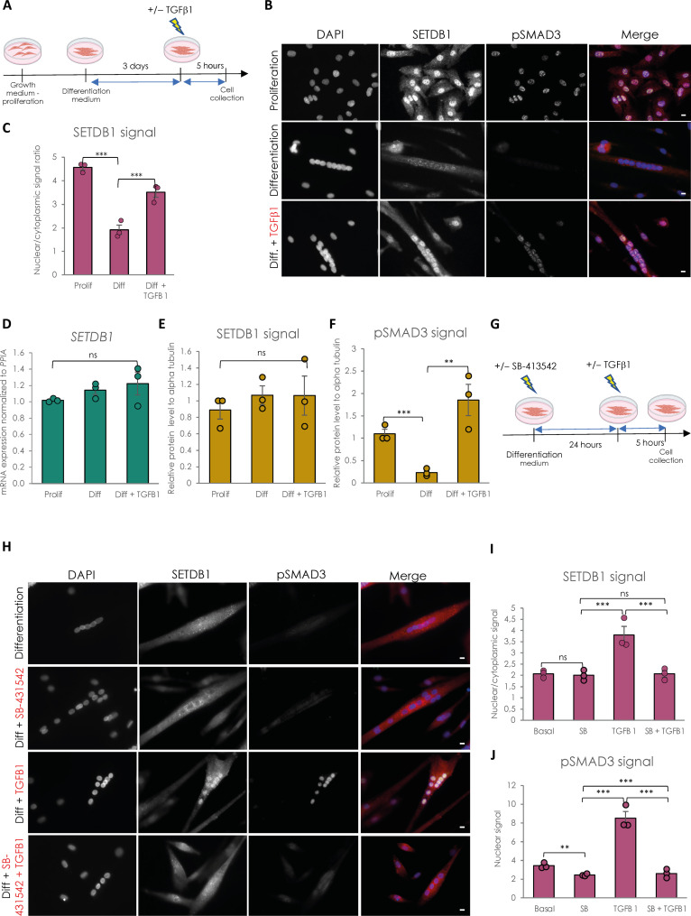
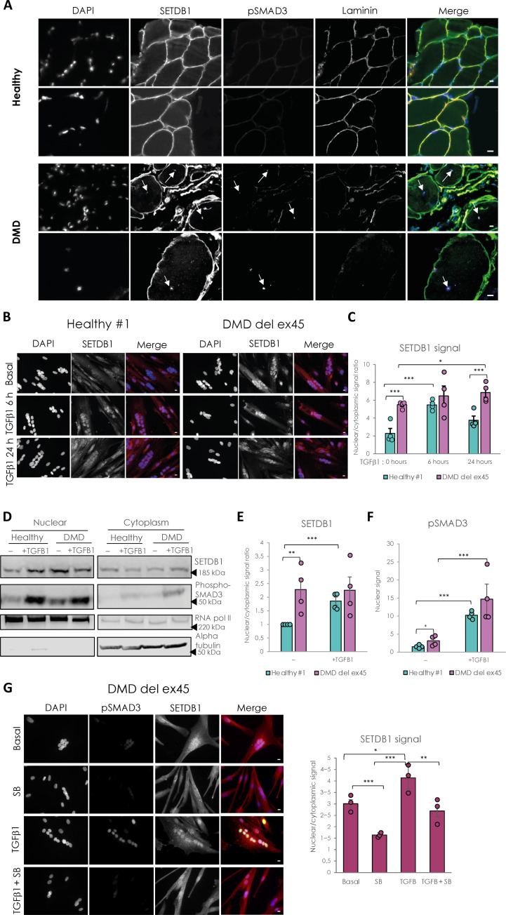
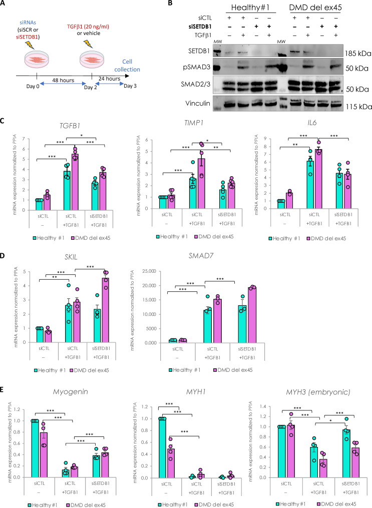
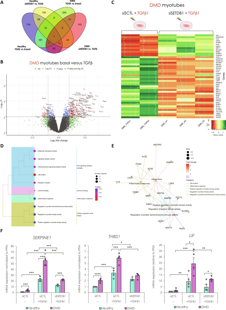
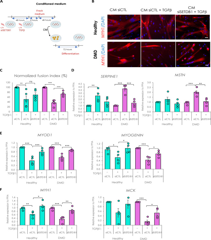
Overactivation of the transforming growth factor-β (TGFβ) signaling in Duchenne muscular dystrophy (DMD) is a major hallmark of disease progression, leading to fibrosis and muscle dysfunction. Here, we investigated the role of SETDB1 (SET domain, bifurcated 1), a histone lysine methyltransferase involved in muscle differentiation. Our data show that, following TGFβ induction, SETDB1 accumulates in the nuclei of healthy myotubes while being already present in the nuclei of DMD myotubes where TGFβ signaling is constitutively activated. Transcriptomics revealed that depletion of SETDB1 in DMD myotubes leads to down-regulation of TGFβ target genes coding for secreted factors involved in extracellular matrix remodeling and inflammation. Consequently, SETDB1 silencing in DMD myotubes abrogates the deleterious effect of their secretome on myoblast differentiation by impairing myoblast pro-fibrotic response. Our findings indicate that SETDB1 potentiates the TGFβ-driven fibrotic response in DMD muscles, providing an additional axis for therapeutic intervention.


Figures

Fig. 1.
Fig. 1.. SETDB1 is excluded from myotube nuclei upon differentiation and can be relocated in response to TGFβ pathway activation.
(A) Scheme of experimental design. Cells were collected in proliferating phase (myoblasts) or after 3 days of differentiation (myotubes) with or without TGFβ1 treatment at 20 ng/ml. (B) Immunostaining of SETDB1 (red) and phospho-SMAD3 (green). Nuclei were stained with 4′,6-diamidino-2-phenylindole (DAPI) (blue). Scale bars, 10 μm. (C) Quantification of SETDB1 nuclear/cytoplasmic signal ratio. (D) Quantitative reverse transcription polymerase chain reaction (RT-qPCR) of SETDB1 in healthy proliferating myoblasts and differentiated myotubes +/− TGFβ1. (E and F) Quantification of SETDB1 and phospho-SMAD3 protein levels measured by Western blot (see fig. S1C) in proliferating myoblasts and myotubes treated or not with TGFβ1 in total protein extracts. Alpha tubulin was used as a loading control to normalize samples. (G) Scheme of experimental design with TGFβ inhibitor SB-431542. (H) Immunostaining of SETDB1 (red) and phospho-SMAD3 (green). Nuclei were stained with DAPI (blue). SB-431542 blocks SETDB1 relocalization in myotube nuclei upon TGFβ1 treatment. Scale bars, 10 μm. (I and J) Quantification of SETDB1 nuclear/cytoplasmic signal ratio (I) and phospho-SMAD3 nuclear signal (J). For all panels, statistics were performed on ≥3 biological replicates (>100 nuclei for immunostaining quantification), and data are represented as average ± SEM. **P < 0.01; ***P < 0.001 (unpaired Student’s t test). ns, not significant.
Fig. 2.
Fig. 2.. DMD-differentiated muscle cells display constitutive relocalization of SETDB1 in the nuclei and higher activation of the TGFβ/SMAD pathway.
(A) Immunostaining of SETDB1 (green), pSMAD3 (magenta), and laminin (red) on histological slides from healthy individual or DMD patient paravertebral muscles. Nuclei were stained with DAPI (blue). White arrows identify centrally localized nuclei of damaged myofibers in the DMD muscle section. Scale bars, 10 μm. (B) Immunostaining of SETDB1 (red) in healthy #1 and DMD del ex45 myotubes. Nuclei were stained with DAPI (blue). Scale bars, 10 μm. (C) Quantification of SETDB1 nuclear/cytoplasmic signal ratio. (D) Western blot of nuclear and cytoplasmic fractions of healthy and DMD myotubes in response to TGFβ1 showing protein levels of SETDB1 and phospho-SMAD3. RNA polymerase II and alpha tubulin were used as loading controls of nuclear and cytoplasmic fractions, respectively. (E and F) Quantification of SETDB1 nuclear/cytoplasmic ratio (E) and phospho-SMAD3 nuclear signal (F). (G) Immunostaining of SETDB1 (red) and phospho-SMAD3 (green) in DMD del ex45 myotubes and quantification of SETDB1 nuclear/cytoplasmic signal ratio. Nuclei were stained with DAPI (blue). Scale bars, 10 μm. For all panels, statistics were performed on ≥3 biological replicates (>100 nuclei for immunostaining quantification), and data are represented as average ± SEM. *P < 0.05; **P < 0.01; ***P < 0.001 (unpaired Student’s t test).
Fig. 3.
Fig. 3.. SETDB1 silencing leads to a decreased response to TGFβ1 in DMD myotubes while key myogenic markers increase.
(A) Scheme of experimental design. Cells were treated with siRNAs scrambled (siSCR) or against SETDB1 for 2 days after 3 days of differentiation (myotubes) and then treated or not with TGFβ1 at 20 ng/ml. (B) Western blot showing SETDB1, phospho-SMAD3, and total SMAD2/3 protein levels. Vinculin was used as a loading control. siCTL, scrambled siRNA (C) RT-qPCR of TGFβ/SMAD pathway known targets TGFβ1, IL6, and TIMP1 in healthy and DMD myotubes +/− siSETDB1 +/− TGFβ1. (D) RT-qPCR of TGFβ/SMAD pathway inhibitors SKIL and SMAD7 in healthy and DMD myotubes +/− siSETDB1 +/− TGFβ1. (E) RT-qPCR of myogenic markers Myogenin, MYH1, and MYH3 (embryonic MHC). For all panels, statistics were performed on ≥3 biological replicates, and data are represented as average ± SEM. *P < 0.05; **P < 0.01; ***P < 0.001 (unpaired Student’s t test).
Fig. 4.
Fig. 4.. SETDB1 silencing in DMD myotubes leads to a decreased expression of TGFβ–dependent genes involved in ECM remodeling, receptor signaling transduction, and inflammation.
(A) Venn diagram shows that a different set of TGFβ1–responsive genes were differentially expressed upon SETDB1 silencing in healthy and DMD myotubes. (B) Volcano plot of differentially expressed genes in DMD myotubes +/− TGFβ1. (C) Heatmap of expression level z-scores for DEGs in DMD myotubes for TGF-β versus TGFβ + siSETDB1 comparison. Some genes found to be increased upon TGFβ1 treatment decrease upon SETDB1 silencing (IGFBP3, THBS1, and IL6ST). (D and E) Enriched categories tree plot (D) and enriched gene-concept network for biological pathways deregulated for TGFβ versus TGFβ + siSETDB1 in DMD myotubes. (F) mRNA levels of gene validated by RT-qPCR involved in ECM remodeling (THBS1 and SERPINE1) and inflammation (LIF). For all panels, statistics were performed on ≥3 biological replicates, and data are represented as average ± SEM. *P < 0.05; **P < 0.01; ***P < 0.001 (unpaired Student’s t test).
Fig. 5.
Fig. 5.. Secretome of SETDB1 deficient myotubes in response to TGFβ1 reduces the negative impact of TGFβ treatment on myoblast differentiation.
(A) Diagram of the conditioned medium experiments. (B) Immunofluorescence of myosin heavy chain (red) and nuclei staining with DAPI (blue). Scale bars, 50 μm. (C) Quantification of fusion index after 6 days of differentiation in the conditioned medium. (D) RT-qPCR of fibrotic markers SERPINE1 and MSTN after 3 days in the conditioned medium. (E) RT-qPCR of early myogenic markers MYOD1 and MYOGENIN. (F) RT-qPCR of late myogenic markers MYH1 and MCK. For all panels, statistics were performed on ≥3 biological replicates (>100 nuclei for immunostaining quantification), and data are represented as average ± SEM. *P < 0.05; **P < 0.01; ***P < 0.001 (unpaired Student’s t test).

Similar articles

Cited by

References

    1. Mozzetta C., Boyarchuk E., Pontis J., Ait-Si-Ali S., Sound of silence: The properties and functions of repressive Lys methyltransferases. Nat. Rev. Mol. Cell Biol. 16, 499–513 (2015). - PubMed
    1. Padeken J., Methot S. P., Gasser S. M., Establishment of H3K9-methylated heterochromatin and its functions in tissue differentiation and maintenance. Nat. Rev. Mol. Cell Biol. 23, 623–640 (2022). - PMC - PubMed
    1. Markouli M., Strepkos D., Piperi C., Structure, activity and function of the SETDB1 protein methyltransferase. Life 11, 817 (2021). - PMC - PubMed
    1. Beyer S., Pontis J., Schirwis E., Battisti V., Rudolf A., Le Grand F., Ait-Si-Ali S., Canonical Wnt signalling regulates nuclear export of Setdb1 during skeletal muscle terminal differentiation. Cell Discov. 2, 16037 (2016). - PMC - PubMed
    1. Juznic L., Peuker K., Strigli A., Brosch M., Herrmann A., Hasler R., Koch M., Matthiesen L., Zeissig Y., Loscher B. S., Nuber A., Schotta G., Neumeister V., Chavakis T., Kurth T., Lesche M., Dahl A., von Massenhausen A., Linkermann A., Schreiber S., Aden K., Rosenstiel P. C., Franke A., Hampe J., Zeissig S., SETDB1 is required for intestinal epithelial differentiation and the prevention of intestinal inflammation. Gut 70, 485–498 (2021). - PMC - PubMed

Publication types

MeSH terms

Substances