. 2024 Sep 2;120(10):1126-1137.
doi: 10.1093/cvr/cvae092.

The ketogenic diet does not improve cardiac function and blunts glucose oxidation in ischaemic heart failure

Affiliations

The ketogenic diet does not improve cardiac function and blunts glucose oxidation in ischaemic heart failure

Kim L Ho et al. Cardiovasc Res. .

Abstract

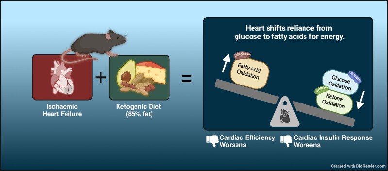
Aims: Cardiac energy metabolism is perturbed in ischaemic heart failure and is characterized by a shift from mitochondrial oxidative metabolism to glycolysis. Notably, the failing heart relies more on ketones for energy than a healthy heart, an adaptive mechanism that improves the energy-starved status of the failing heart. However, whether this can be implemented therapeutically remains unknown. Therefore, our aim was to determine if increasing ketone delivery to the heart via a ketogenic diet can improve the outcomes of heart failure.

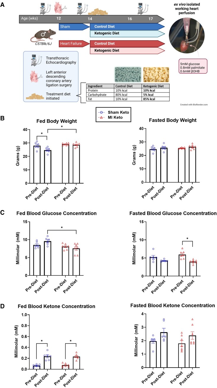
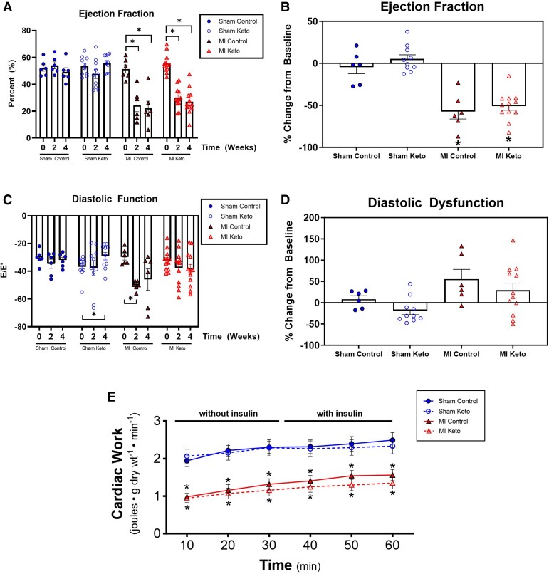
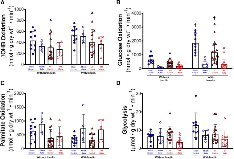
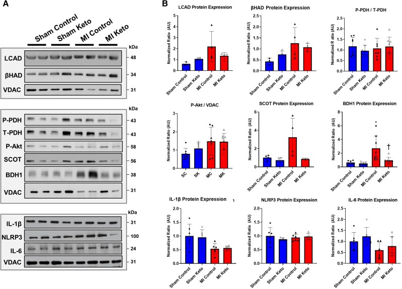
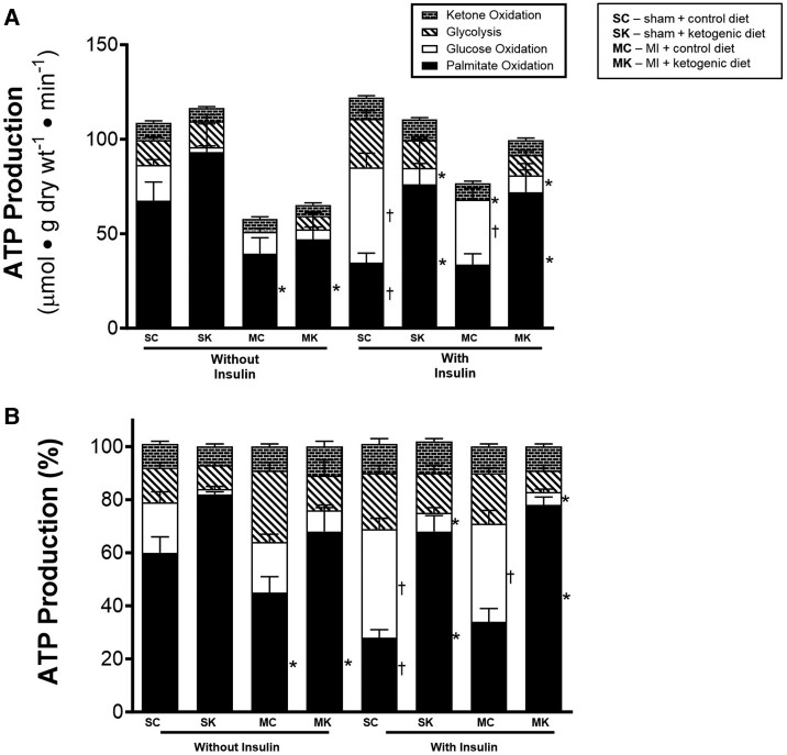
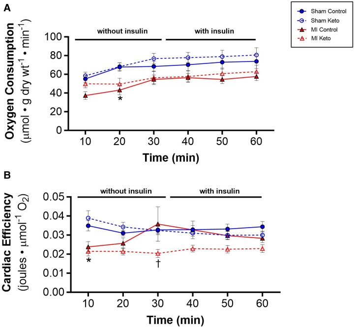
Methods and results: C57BL/6J male mice underwent either a sham surgery or permanent left anterior descending coronary artery ligation surgery to induce heart failure. After 2 weeks, mice were then treated with either a control diet or a ketogenic diet for 3 weeks. Transthoracic echocardiography was then carried out to assess in vivo cardiac function and structure. Finally, isolated working hearts from these mice were perfused with appropriately 3H or 14C labelled glucose (5 mM), palmitate (0.8 mM), and β-hydroxybutyrate (β-OHB) (0.6 mM) to assess mitochondrial oxidative metabolism and glycolysis. Mice with heart failure exhibited a 56% drop in ejection fraction, which was not improved with a ketogenic diet feeding. Interestingly, mice fed a ketogenic diet had marked decreases in cardiac glucose oxidation rates. Despite increasing blood ketone levels, cardiac ketone oxidation rates did not increase, probably due to a decreased expression of key ketone oxidation enzymes. Furthermore, in mice on the ketogenic diet, no increase in overall cardiac energy production was observed, and instead, there was a shift to an increased reliance on fatty acid oxidation as a source of cardiac energy production. This resulted in a decrease in cardiac efficiency in heart failure mice fed a ketogenic diet.

Conclusion: We conclude that the ketogenic diet does not improve heart function in failing hearts, due to ketogenic diet-induced excessive fatty acid oxidation in the ischaemic heart and a decrease in insulin-stimulated glucose oxidation.

Keywords: Cardiac energy metabolism; Ischaemic heart failure; Isolated working heart perfusion; Ketogenic diet; Metabolism.


Conflict of interest statement

Conflict of interest: none declared.

Figures

Graphical Abstract
Graphical Abstract
Figure 1
Figure 1
Experimental protocol and effect of the ketogenic diet on body weight, blood glucose, and blood ketones. (A) Experimental protocol. Please see methods for specific details. (B) Fed and fasted body weight of mice pre- and post-control/ketogenic diet implementation. (C) Fed and fasted blood glucose of mice pre- and post-control/ketogenic diet implementation. (D) Fed and fasted β-OHB levels of mice pre- and post-control/ketogenic diet implementation. Sham keto mice n = 6; MI keto mice n = 7. Values are the mean ± SEM. *P < 0.05 compared with the pre-dietary implementation experimental group (two-way multiple comparisons ANOVA with Sidak post hoc test).
Figure 2
Figure 2
Ketogenic diet effects on in vivo and ex vivo cardiac function in post-MI mice. Transthoracic echocardiographic measurements of in vivo cardiac function: (A, B) ejection fraction and (C, D) diastolic function. Sham control mice n = 6; MI control mice n = 6; sham keto mice n = 10; MI keto mice n = 12. (E) Ex vivo measurement of cardiac function in isolated working hearts. Sham control mice n = 19; MI control mice n = 25; sham keto mice n = 11; MI keto mice n = 16. Values are the mean ± SEM. *P < 0.05 compared with the respective sham group (one-way multiple comparisons ANOVA with Sidak post hoc test for A–D; two-way multiple comparisons ANOVA with Sidak post hoc test for E).
Figure 3
Figure 3
Ketogenic diet effects on β-OHB oxidation, glucose oxidation, fatty acid oxidation, and glycolysis in post-MI hearts. Cardiac energy metabolic rates were measured in isolated working hearts: (A) β-OHB oxidative rates without and with insulin (ketone body oxidation rates), (B) glucose oxidation rates without and with insulin, (C) palmitate oxidation rates without and with insulin, and (D) glycolytic rates without and with insulin. For A and C, sham control mice n = 10; MI control mice n = 16; sham keto mice n = 5; and MI keto mice n = 6. For B and D, sham control mice n = 9; MI control mice n = 16; sham keto mice n = 5; and MI keto mice n = 11. Values are the mean ± SEM. *P < 0.05 compared with respective control group, †P < 0.05 compared with the ‘without insulin’ group (two-way multiple comparisons ANOVA with Sidak post hoc test).
Figure 4
Figure 4
Ketogenic diet effects on cardiac protein expression of various metabolic and inflammatory targets in post-MI hearts: (A, B) LCAD, β-HAD, phospho-pyruvate dehydrogenase (p-PDH), serine/threonine protein kinase Akt (Akt), SCOT, BDH1, IL-1β, NACHT, LRR, PYD domains containing protein 3 (NLRP3) inflammasome, and IL-6. Sham control mice n = 7; MI control mice n = 8; sham keto mice n = 8; and MI keto mice n = 8. Values are the mean ± SEM. *P < 0.05 compared with sham control (two-way multiple comparisons ANOVA with Sidak post hoc test).
Figure 5
Figure 5
Ketogenic diet effects on cardiac ATP production rates in post-MI hearts. (A) Absolute ATP production rates coming from palmitate oxidation, glucose oxidation, ketone oxidation, and glycolysis without and with insulin. (B) Per cent contribution of palmitate oxidation, glucose oxidation, ketone oxidation, and glycolysis to total per cent of ATP production without and with insulin. For palmitate and ketone oxidation: sham control mice n = 10; MI control mice n = 16; sham keto mice n = 5; and MI keto mice n = 6. For glycolysis and glucose oxidation: sham control mice n = 9; MI control mice n = 16; sham keto mice n = 5; and MI keto mice n = 13. Values are the mean ± SEM. P < 0.05 compared with respective control group, †P < 0.05 compared with the ‘without insulin’ group (two-way multiple comparisons ANOVA with Sidak post hoc test).
Figure 6
Figure 6
Ketogenic diet on oxygen consumption and cardiac efficiency in post-MI hearts. (A) Oxygen consumption rates as measured in isolated working hearts. (B) Cardiac efficiency as determined by normalizing ex vivo cardiac work to oxygen consumption rates. Sham control mice n = 16; MI control mice n = 22; sham keto mice n = 11; and MI keto mice n = 12. Values are the mean ± SEM. *P < 0.05 compared with the respective sham group, †P < 0.05 compared with respective control diet group (two-way multiple comparisons ANOVA with Sidak post hoc test).

Comment in

Similar articles

Cited by

References

    1. Emmons-Bell S, Johnson C, Roth G. Prevalence, incidence and survival of heart failure: a systematic review. Heart 2022;108:1351–1360. - PMC - PubMed
    1. Bing RJ. The metabolism of the heart. Harvey Lect 1954;50:27–70. - PubMed
    1. Neubauer S, Horn M, Cramer M, Harre K, Newell John B, Peters W, Pabst T, Ertl G, Hahn D, Ingwall Joanne S, Kochsiek K. Myocardial phosphocreatine-to-ATP ratio is a predictor of mortality in patients with dilated cardiomyopathy. Circulation 1997;96:2190–2196. - PubMed
    1. Lopaschuk GD, Karwi QG, Tian R, Wende AR, Abel ED. Cardiac energy metabolism in heart failure. Circ Res 2021;128:1487–1513. - PMC - PubMed
    1. Karwi QG, Uddin GM, Ho KL, Lopaschuk GD. Loss of metabolic flexibility in the failing heart. Front Cardiovasc Med 2018;5:68. - PMC - PubMed

MeSH terms