. 2024 Apr 30;15(1):3666.
doi: 10.1038/s41467-024-47963-5.

Airway epithelial CD47 plays a critical role in inducing influenza virus-mediated bacterial super-infection

Affiliations

Airway epithelial CD47 plays a critical role in inducing influenza virus-mediated bacterial super-infection

Sungmin Moon et al. Nat Commun. .

Abstract

Respiratory viral infection increases host susceptibility to secondary bacterial infections, yet the precise dynamics within airway epithelia remain elusive. Here, we elucidate the pivotal role of CD47 in the airway epithelium during bacterial super-infection. We demonstrated that upon influenza virus infection, CD47 expression was upregulated and localized on the apical surface of ciliated cells within primary human nasal or bronchial epithelial cells. This induced CD47 exposure provided attachment sites for Staphylococcus aureus, thereby compromising the epithelial barrier integrity. Through bacterial adhesion assays and in vitro pull-down assays, we identified fibronectin-binding proteins (FnBP) of S. aureus as a key component that binds to CD47. Furthermore, we found that ciliated cell-specific CD47 deficiency or neutralizing antibody-mediated CD47 inactivation enhanced in vivo survival rates. These findings suggest that interfering with the interaction between airway epithelial CD47 and pathogenic bacterial FnBP holds promise for alleviating the adverse effects of super-infection.


Conflict of interest statement

The authors declare no competing interests.

Figures
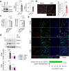
Fig. 1
Fig. 1. Influenza virus induces CD47 expression on the apical surface of ciliated cells in an NF-κB-dependent manner.
HBECs were infected with (+ IAV) or without (Mock) influenza A virus. a Immunoblot analysis of junction protein ZO-1 and surface protein CD47 at 1 day post-infection (dpi). Normalized CD47 and ZO-1 protein levels are presented as bar graphs (Mock, n = 4; + IAV, n = 4). b Representative whole-mount images of ZO-1 (white) and CD47 (red) at 1 dpi. The area where ZO-1 disconnection occurred are presented as bar graphs (Mock, n = 6; + IAV, n = 6). c Quantitative PCR (qPCR) analysis of ZO-1 and CD47 mRNAs at 1 and 3 dpi [Mock, n = 3; + IAV (1 dpi), n = 3; + IAV (3 dpi), n = 3]. d HBECs were treated with or without 10 μM NF-κB inhibitor caffeic acid phenethyl ester (CAPE) for 1 h before influenza virus infection. Immunoblot analysis of ICAM-1 and CD47 at 1 dpi. Normalized ICAM-1 and CD47 protein levels are presented as bar graphs (DMSO + Mock, n = 4; DMSO + IAV, n = 4; CAPE + Mock, n = 4; CAPE + IAV, n = 4). e Whole-mount images of influenza virus-infected HBECs. Co-staining of CD47 (red) and ciliated cell-specific marker protein Ac-α-tubulin (green, n = 4) or goblet cell-specific marker protein MUC5AC (cyan, n = 3). Percentages of CD47-positive cells are presented as bar graphs. Data are presented as mean values ± SEM. Significance was determined by unpaired two-tailed Student’s t test or one-way ANOVA with Tukey’s multiple comparisons test. n.s. not significant. Source data are provided as a Source Data file.
Fig. 2
Fig. 2. Knock-down or neutralization of CD47 protects HBECs from viral–bacterial co-infection.
For CD47 knock-down, HBECs were transfected with scrambled shRNA (sc-shRNA) or shRNA targeting CD47 (CD47 shRNA) using a lentiviral delivery system. For CD47 neutralization, HBECs were treated with either IgG1 (MOPC-21) or α-hCD47 (B6H12.2) antibodies. a Gene expression of CD47 was analyzed using qRT-PCR (normalized by GAPDH mRNA) (sc-shRNA, n = 4; CD47 shRNA, n = 4). b Protein expression of CD47 was analyzed using immunoblotting (sc-shRNA, n = 5; CD47 shRNA, n = 5). c Paracellular FITC-dextran permeability was measured in Mock (n = 16), Virus only (IAV, n = 16), Bacteria only (S. aureus, n = 16), sc-shRNA + Super-infection (n = 12), CD47 shRNA + Super-infection (n = 6), IgG1 + Super-infection (n = 6), and α-hCD47 antibodies (Ab) + Super-infection (n = 8). d Trans-epithelial resistance was measured in Mock (n = 15), IAV (n = 14), S. aureus (n = 13), sc-shRNA + Super-infection (n = 13), CD47 shRNA + Super-infection (n = 6), IgG1 + Super-infection (n = 6), and α-hCD47 Ab + Super-infection (n = 8). e Microscopic images of HBECs at 5 dpi. The percentage of the damaged area is presented as bar graphs (Mock, n = 3; Virus only, n = 3; Bacteria only, n = 3; Super-infection, n = 4; IgG1 + Super-infection, n = 4; α-hCD47 Ab + Super-infection, n = 4). Data are presented as mean values ± SEM. Significance was determined by unpaired two-tailed Student’s t test or one-way ANOVA with Tukey’s multiple comparisons test. N.D. not determined. Source data are provided as a Source Data file.
Fig. 3
Fig. 3. Epithelial CD47 and bacterial FnBP directly interacts.
a, b Influenza virus-infected HBECs (IAV) infected with live S. aureus or S. aureus-derived samples (S, supernatant of S. aureus-cultured media; U, UV-killed S. aureus; H, heat-killed S. aureus). Paracellular FITC-dextran permeability (a) and trans-epithelial electrical resistance (b) of 7 groups: i) Mock (n = 3), ii) Virus only (n = 3), iii) Bacteria only (n = 3), iv) Super-infection (n = 3), v) Virus with S (n = 3), vi) Virus with U (n = 3), and vii) Virus with H (n = 3). c Whole mount image of CD47 (red) and S. aureus (green) in the influenza virus-infected HBECs at 1 dpi. An open arrow head indicates CD47+ cell without S. aureus and closed arrow heads indicate CD47+ cells with S. aureus. Co-localization of CD47+ cells and S. aureus are presented as violin plots (Mock, n = 6; + IAV, n = 6). dg Bacterial adhesion assay. Colonization of S. aureus was assessed in HBECs inoculated with the virus (MOI 1) for 2 h before treatment with IgG1 control antibodies or α-hCD47 neutralizing antibodies (2 h), followed by S. aureus (MOI 3) infection for 3 h (IgG1 + S. aureus, n = 4; α-hCD47 Ab + S. aureus, n = 4; IgG1 + Super-infection, n = 4; α-hCD47 Ab+ Super-infection, n = 4) (d). Adherence of S. aureus WT (FnBP A+/B+, n = 4) and three mutant strains (FnBP A+/B–, n = 4; FnBP A–/B+, n = 4; FnBP A–/B–, n = 4) was assessed in HBECs inoculated with the virus (MOI 1) for 2 h, followed by S. aureus (MOI 3) infection for 3 h (e). Colonization of S. aureus WT (FnBP A+/B+) (f) and double deletion mutant (FnBP A–/B–) (g) was assessed in HBECs inoculated with the virus (MOI 1) for 2 h before treatment with IgG1 control antibodies (n = 4) or α-hCD47 neutralizing antibodies (1, 5, and 10 μg/mL for FnBP A+/B+, and 10 μg/mL for FnBP A–/B–, n = 4 each) for 2 h, followed by S. aureus (MOI 3) infection for 3 h. h Pull-down assay using His-tagged hCD47 recombinant protein. Bacterial plating [FnBP A+/B+ and FnBP A–/B– (n = 3 each in the absence or in the presence of His-tagged hCD47)] and immunoblot analysis were performed using supernatants and pellets after separation with α-His-Dynabeads™/DynaMag™−2 system. The graphs present the percentage of colony numbers grown in the culture of the supernatants or the pellets. Data are presented as mean values ± SEM. Significance was determined by one-way ANOVA with Tukey’s multiple comparisons test or unpaired two-tailed Student’s t test. n.s. not significant. Source data are provided as a Source Data file.
Fig. 4
Fig. 4. Foxj1Cre-Specific, but not LysMCre-Specific, CD47 disruption protects mice from super-infection.
6–8-weeks-old (18–21 g of body weight) FoxJ1-Cre;floxed (CD47Foxj1), LysM-Cre;floxed (CD47LysM), and control floxed (CD47f/f) mice were infected with 100 PFU of influenza virus on day 0, and 1 ×10⁸ CFU of S. aureus on day 7. abkl Body weight loss (a, k) and survival rates (b, l) were monitored for 29 days. The dotted line indicates the body weight exclusion cut-off. A mantel cox survival analysis was used to compare the survival rates between groups. The numbers in parenthesis represent the count of surviving mice. cm Representative hematoxylin and eosin (H&E) staining of lung sections. The dotted lines indicate lymphocytic infiltration and arrows indicate alveolar hemorrhage. Lung injury scores are presented as violin plots in CD47Foxj1 mice (CD47f/f, n = 8; CD47Foxj1 n = 5) (c) and CD47LysM mice (CD47f/f, n = 8; CD47LysM, n = 8) (m). dj, nt Tissue injury parameters were measured at 24 h after bacterial infection; total cell number in BAL fluids (BALF) of CD47Foxj1 mice (CD47f/f, n = 5; CD47Foxj1, n = 5) (d) and CD47LysM mice (CD47f/f, n = 8; CD47LysM, n = 10) (n); bacterial adherence in the lung of CD47Foxj1 mice (CD47f/f, n = 10; CD47Foxj1, n = 11) (e) and CD47LysM mice (CD47f/f, n = 13; CD47LysM, n = 15) (o); bacterial invasion in the lung of CD47Foxj1 mice (CD47f/f, n = 7; CD47Foxj1, n = 7) (f) and CD47LysM mice (CD47f/f, n = 5; CD47LysM, n = 5) (p); and bacterial burden in the spleen of CD47Foxj1 mice (CD47f/f, n = 5; CD47Foxj1, n = 6) (g) and CD47LysM mice (CD47f/f, n = 5; CD47LysM, n = 5) (q); total protein concentrations in BALF of CD47Foxj1 mice (CD47f/f, n = 5; CD47Foxj1, n = 5) (h) and CD47LysM mice (CD47f/f, n = 8; CD47LysM, n = 10) (r); and inflammatory cytokines TNF-α in BALF of CD47Foxj1 mice (CD47f/f, n = 5; CD47Foxj1, n = 5) (i) and CD47LysM mice (CD47f/f, n = 8; CD47LysM, n = 10) (s), and IL-6 in BALF of CD47Foxj1 mice (CD47f/f, n = 5; CD47Foxj1, n = 5) (j) and CD47LysM mice (CD47f/f, n = 8; CD47LysM, n = 10) (t). Data are presented as mean values ± SEM. Significance was determined by unpaired two-tailed Student’s t test. Source data are provided as a Source Data file.
Fig. 5
Fig. 5. CD47 neutralization enhances protection against super-infection.
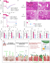
For neutralization test, 6–8-weeks-old (18–21 g of body weight) C57BL/6 WT mice were infected with 10 PFU of influenza virus on day 0 and 5 × 10⁵ CFU of S. aureus on day 7. Before bacterial infection, mice were intranasally treated twice at day 5 and 7 with IgG2a control antibodies (2A3, n = 17) or α-mCD47 neutralizing antibodies (MIAP301, n = 16). a, b Body weight loss (a) and survival rates (b) were monitored for 29 days. The dotted line indicates the body weight exclusion cut-off. A mantel cox survival analysis was used to compare the survival rates between groups. The numbers in parenthesis are numbers of survived mice. c Representative hematoxylin and eosin (H&E) staining of lung sections (IgG2a + Mock, n = 4; α-mCD47 Ab + Mock, n = 4; IgG2a + Virus only, n = 7; α-mCD47 Ab + Virus only, n = 7; IgG2a + Bacteria only, n = 4; α-mCD47 Ab + Bacteria only, n = 4; IgG2a + Super-infection, n = 9; α-mCD47 Ab + Super-infection, n = 11). The dotted lines indicate lymphocytic infiltration and arrows indicate alveolar hemorrhage. Lung injury scores are presented as violin plots. dj Tissue injury parameters were measured at 24 h after bacterial infection (IgG2a + Mock, n = 4; α-mCD47 Ab + Mock, n = 4; IgG2a + Virus only, n = 7; α-mCD47 Ab + Virus only, n = 7; IgG2a + Bacteria only, n = 4; α-mCD47 Ab + Bacteria only, n = 4; IgG2a + Super-infection, n = 7-9; α-mCD47 Ab + Super-infection, n = 7-11); total cell number in BAL fluids (BALF) (d), bacterial adherence (e) and invasion (f) in the lung, and the bacterial burden in the spleen (g), total protein concentrations (h) in BALF, and inflammatory cytokines TNF-α (i) and IL-6 (j) in BALF. Data are presented as mean values ± SEM. Significance was determined by unpaired two-tailed t test. n.s. not significant. N.D. not determined. Source data are provided as a Source Data file.
Fig. 6
Fig. 6. Interaction between epithelial CD47 and bacterial FnBP is essential to cause super-infection.
aj 6–8-weeks-old (18–21 g of body weight) FoxJ1-Cre; floxed (Cd47Foxj1) and control floxed (Cd47f/f) mice were infected with 100 PFU of influenza virus on day 0, and 1 ×10⁸ CFU of S. aureus WT (FnBP A+/B+) and double deletion mutant (FnBP A–/B–) on day 7 (Cd47f/f + FnBP A+/B+, n = 9; Cd47f/f + FnBP A–/B–, n = 11; Cd47Foxj1 + FnBP A+/B+, n = 12; Cd47Foxj1 +FnBP A–/B–, n = 10). Body weight loss (a) and survival rates (b) were monitored for 29 days. The dotted line indicates the body weight exclusion cut-off. A mantel cox survival analysis was used to compare the survival rates between groups. The numbers within circles or squares represent the count of surviving mice. Representative hematoxylin and eosin (H&E) staining of lung sections (c). Lung injury scores are presented as violin plots (Cd47f/f + FnBP A+/B+, n = 4; Cd47f/f + FnBP A–/B–, n = 5; Cd47Foxj1 + FnBP A+/B+, n = 5; Cd47Foxj1 +FnBP A–/B–, n = 5). Tissue injury parameters were measured at 24 h after bacterial infection (Cd47f/f + FnBP A+/B+, n = 4; Cd47f/f + FnBP A–/B–, n = 5; Cd47Foxj1 + FnBP A+/B+, n = 5; Cd47Foxj1 +FnBP A–/B–, n = 5); total cell number in BAL fluids (BALF) (d), bacterial adherence (e) and invasion (f) in the lung, and bacterial burden in the spleen (g), total protein concentrations in BALF (h), and inflammatory cytokines TNF-α (i) and IL-6 (j) in BALF. k Proposed model of viral infection-induced CD47-mediated secondary bacterial infection. Data are presented as mean values ± SEM. Significance was determined by one-way ANOVA with Tukey’s multiple comparisons test. Source data are provided as a Source Data file.

References

    1. Morens DM, Taubenberger JK, Fauci AS. Predominant role of bacterial pneumonia as a cause of death in pandemic influenza: implications for pandemic influenza preparedness. J. Infect. Dis. 2008;198:962–970. doi: 10.1086/591708. - DOI - PMC - PubMed
    1. Estenssoro E, et al. Pandemic 2009 influenza A in Argentina: a study of 337 patients on mechanical ventilation. Am. J. Respir. Crit. Care Med. 2010;182:41–48. doi: 10.1164/201001-0037OC. - DOI - PubMed
    1. Rice TW, et al. Critical illness from 2009 pandemic influenza A virus and bacterial coinfection in the United States. Crit. Care Med. 2012;40:1487–1498. doi: 10.1097/CCM.0b013e3182416f23. - DOI - PMC - PubMed
    1. McCullers JA. The co-pathogenesis of influenza viruses with bacteria in the lung. Nat. Rev. Microbiol. 2014;12:252–262. doi: 10.1038/nrmicro3231. - DOI - PubMed
    1. Georas SN, Rezaee F. Epithelial barrier function: at the front line of asthma immunology and allergic airway inflammation. J. Allergy Clin. Immunol. 2014;134:509–520. doi: 10.1016/j.jaci.2014.05.049. - DOI - PMC - PubMed

Publication types

MeSH terms