. 2024 May 2;73(6):113.
doi: 10.1007/s00262-024-03695-5.

Neutrophil-activating secretome characterizes palbociclib-induced senescence of breast cancer cells

Affiliations

Neutrophil-activating secretome characterizes palbociclib-induced senescence of breast cancer cells

Gabriele Favaretto et al. Cancer Immunol Immunother. .

Abstract

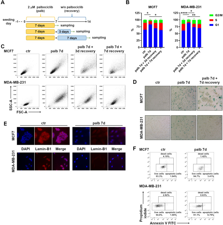
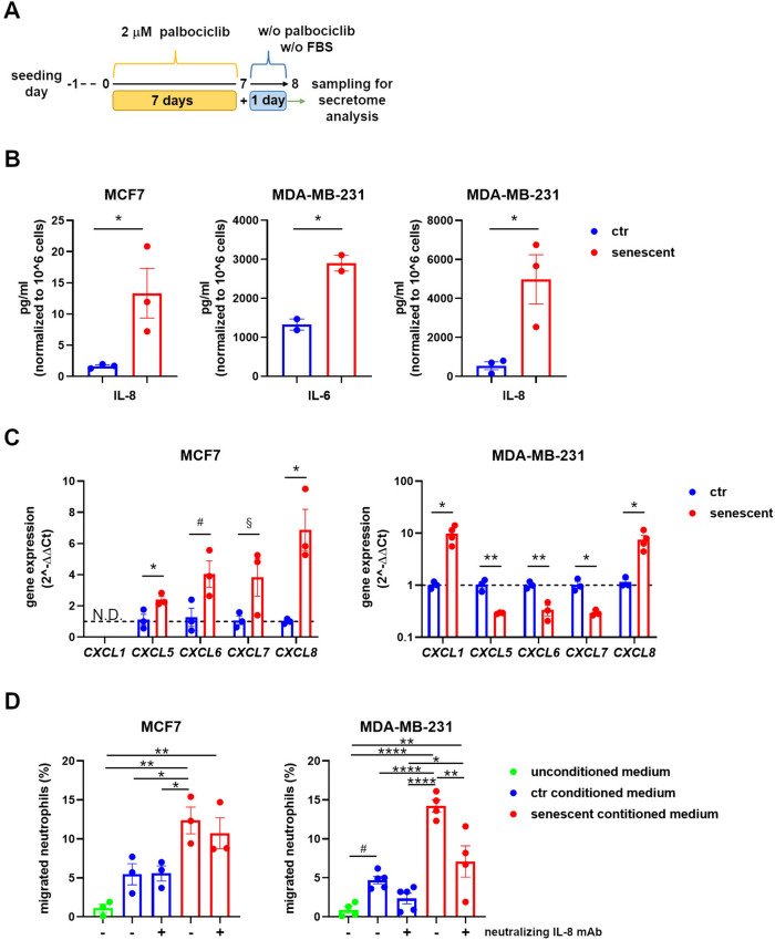
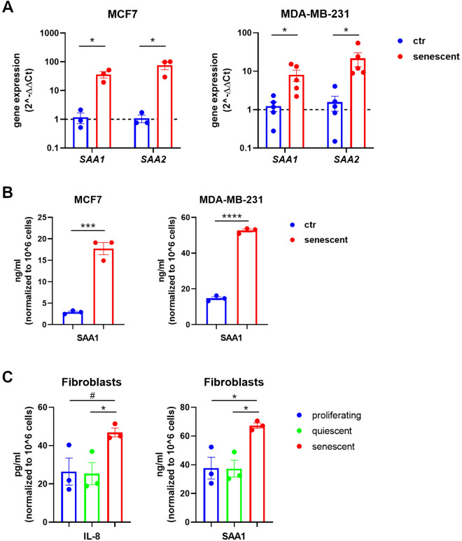
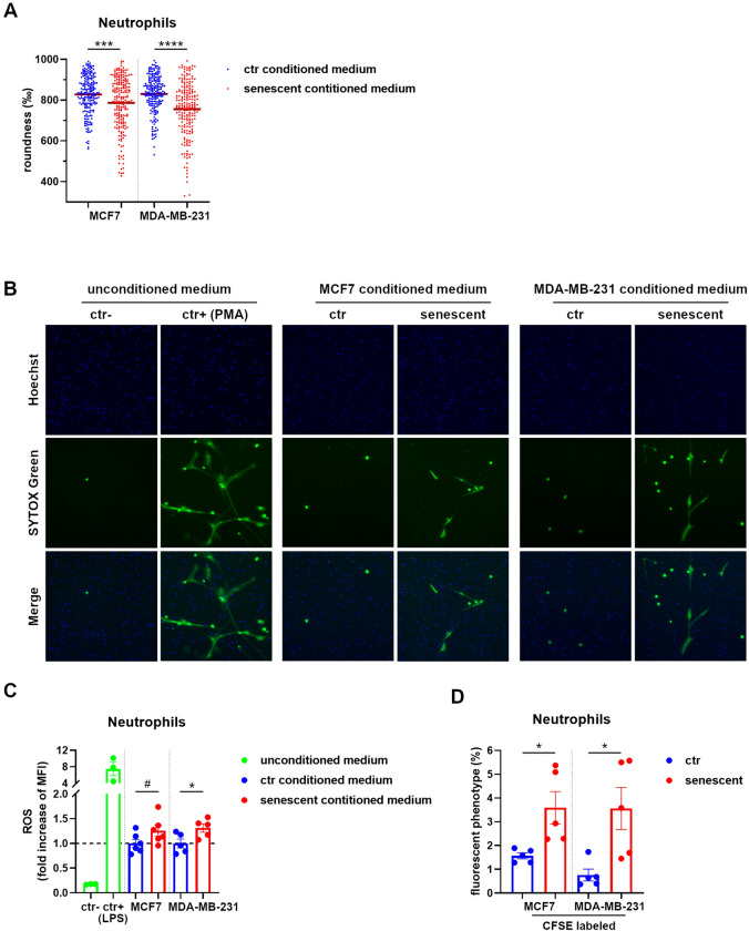
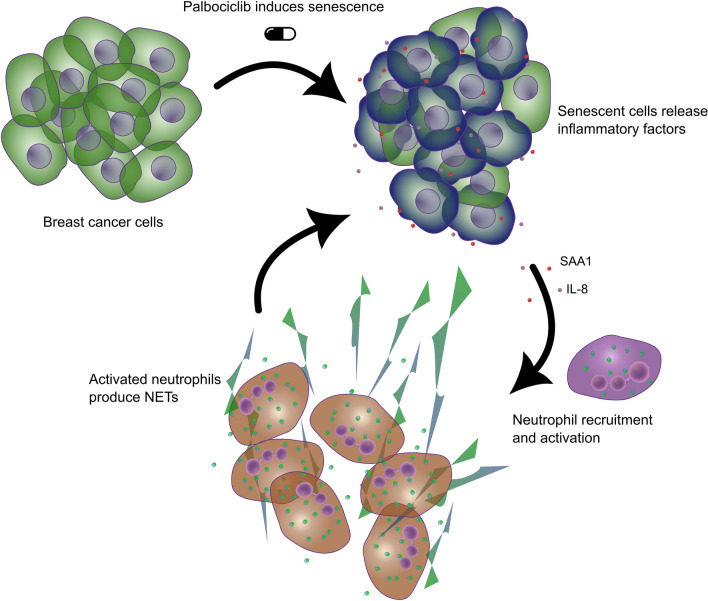
Senescent cells have a profound impact on the surrounding microenvironment through the secretion of numerous bioactive molecules and inflammatory factors. The induction of therapy-induced senescence by anticancer drugs is known, but how senescent tumor cells influence the tumor immune landscape, particularly neutrophil activity, is still unclear. In this study, we investigate the induction of cellular senescence in breast cancer cells and the subsequent immunomodulatory effects on neutrophils using the CDK4/6 inhibitor palbociclib, which is approved for the treatment of breast cancer and is under intense investigation for additional malignancies. Our research demonstrates that palbociclib induces a reversible form of senescence endowed with an inflammatory secretome capable of recruiting and activating neutrophils, in part through the action of interleukin-8 and acute-phase serum amyloid A1. The activation of neutrophils is accompanied by the release of neutrophil extracellular trap and the phagocytic removal of senescent tumor cells. These findings may be relevant for the success of cancer therapy as neutrophils, and neutrophil-driven inflammation can differently affect tumor progression. Our results reveal that neutrophils, as already demonstrated for macrophages and natural killer cells, can be recruited and engaged by senescent tumor cells to participate in their clearance. Understanding the interplay between senescent cells and neutrophils may lead to innovative strategies to cope with chronic or tumor-associated inflammation.

Keywords: Breast cancer; NET; Neutrophil; Palbociclib; SASP; Senescence; Serum amyloid A.


Conflict of interest statement

The authors have no conflicts of interest to declare that are relevant to the content of this article.

Figures

Fig. 1
Fig. 1
Palbociclib induces reversible senescence in breast cancer cell lines. A Diagram depicting the experimental design. B Cell cycle analysis of MCF7 and MDA-MB-231 cells treated with 2 μM palbociclib as outlined in Fig. 1A. Bars show mean ± SEM from three independent flow cytometric experiments. Statistical analysis was performed using two-way ANOVA followed by Tukey’s multiple-comparisons test. *P < 0.05, **P < 0.01, ****P < 0.0001 comparing the G1 phase. C MCF7 and MDA-MB-231 cell morphology after palbociclib treatment as evaluated by forward and side scatter flow cytometric parameters. D Senescence-associated β-galactosidase (SA-β-Gal) assay of MCF7 and MDA-MB-231 cells after one week of 2 μM palbociclib and a further week without palbociclib. Blue staining marks senescent cells. Images are representative of more than three independent experiments. Magnification 200 ×. E Representative immunofluorescence images of DAPI and Lamin-B1 staining of MCF7 and MDA-MB-231 cells treated with 2 μM palbociclib for 1 week. Magnification 400 ×. F Cell death and apoptosis of MCF7 and MDA-MB-231 cells upon palbociclib treatment as evaluated by Annexin V assay. Representative dot plots of two independent experiments
Fig. 2
Fig. 2
Senescent MCF7 and MDA-MB-231 cells recruit neutrophils. A Diagram depicting the experimental design for conditioned medium analysis. B Quantification by ELISA of IL-6 and IL-8 secreted by control proliferating and senescent MCF7 and MDA-MB-231 cells in 24 h-conditioned media as outlined in Fig. 2A. Bars show mean ± SEM and statistical significance was determined by unpaired two-tailed Student’s t-test. *P < 0.05. C Quantitative real-time PCR of the indicated CXCR2 ligands from total RNA extracted from control proliferating and senescent MCF7 and MDA-MB-231 cells. Data are reported as fold increase by using the 2−ΔΔCt method and the average of control samples for the ΔΔ calculation. Bars show mean ± SEM from at least three independent experiments. Statistical analysis was performed using multiple t-test (one for each gene of interest). *P < 0.05, **P < 0.01, #P = 0,055, §P = 0,092. N.D. = not detected. D Percentage of neutrophils migrated toward conditioned media obtained from control proliferating and senescent MCF7 and MDA-MB-231 cells. Migration toward medium alone (unconditioned medium) was considered as background. Experiments were performed both in the absence and in the presence of neutralizing IL-8 mAb as indicated. Bars show mean ± SEM from at least three independent experiments. Statistical analysis was performed using one-way ANOVA followed by Tukey’s multiple-comparisons test. *P < 0.05, **P < 0.01, ****P < 0.0001, #P = 0.087
Fig. 3
Fig. 3
Senescent cells express recruiting and activating factors for neutrophils. A Quantitative real-time PCR of SAA1 and SAA2 genes from total RNA extracted from control proliferating and senescent MCF7 and MDA-MB-231 cells. Data are reported as fold increase by using the 2−ΔΔCt method and the average of control samples for the ΔΔ calculation. Bars show mean ± SEM from at least three independent experiments. Statistical analysis was performed using multiple t-test (one for each gene of interest). *P < 0.05. B Quantification by ELISA of SAA1 secreted by control proliferating and senescent MCF7 and MDA-MB-231 cells in 24 h-conditioned media. Bars show mean ± SEM and statistical significance was determined by unpaired two-tailed Student’s t-test. ***P < 0.001, ****P < 0.0001. C Quantification by ELISA of IL-8 and SAA1 secreted by proliferating, quiescent, and senescent human foreskin fibroblasts in 24 h-conditioned media. Bars show mean ± SEM and statistical significance was determined by one-way ANOVA followed by Tukey’s multiple-comparisons test. *P < 0.05, #P = 0.091
Fig. 4
Fig. 4
Secretome of senescent MCF7 and MDA-MB-231 cells promotes neutrophil activation. A Circularity score of neutrophils stimulated with the conditioned media obtained from control proliferating and senescent MCF7 and MDA-MB-231 cells. Cell shape was visualized by phalloidin staining and roundness calculated with the ImageJ software. Each dot represents the corresponding value (1000 indicates precise round shape) of a single cell. The horizontal red bar represents the mean value. Statistical analysis was performed using unpaired two-tailed Student’s t-test (one for each cell line). ***P < 0.001, ****P < 0.0001. B NET formation by neutrophils stimulated with the conditioned media obtained from control proliferating and senescent MCF7 and MDA-MB-231 cells. Medium alone (with FBS) and medium with PMA were used as negative and positive control, respectively. Nuclei and NETs were visualized by Hoechst and SYTOX Green staining, respectively. Images are representative of two independent experiments. Magnification 100 × . C ROS production by neutrophils stimulated with the conditioned media obtained from control proliferating and senescent MCF7 and MDA-MB-231 cells. Medium alone (with FBS) and medium with LPS were used as negative and positive control, respectively. ROS were quantified by flow cytometry considering the median fluorescence intensity (MFI) of ROS-specific fluorogenic probes. Data are reported as fold increase in respect to the average of corresponding  control proliferating samples. Bars show mean ± SEM from at least three independent experiments. Statistical analysis was performed using unpaired two-tailed Student’s t-test (one for each cell line). *P < 0.05, #P = 0.088. D Evaluation of the uptake by neutrophils of CFSE-labeled control proliferating or senescent MCF7 and MDA-MB-231 cells as evaluated by flow cytometry. Graphs show the percentage of neutrophils acquiring the CFSE+ phenotype. Bars show mean ± SEM from at least three independent experiments. Statistical analysis was performed using unpaired two-tailed Student’s t-test (one for each cell line). *P < 0.05
Fig. 5
Fig. 5
Senescence by palbociclib recruits and activates neutrophils. Breast cancer cells induced to senescence by the CDK4/6 inhibitor palbociclib release IL-8 and SAA1, which, in turn, recruit and activate neutrophils causing NET production in tumor

References

    1. Galluzzi L, Vitale I, Aaronson SA, et al. Molecular mechanisms of cell death: recommendations of the Nomenclature Committee on cell death 2018. Cell Death Differ. 2018;25:486–541. doi: 10.1038/s41418-017-0012-4. - DOI - PMC - PubMed
    1. Gluck S, Guey B, Gulen MF, et al. Innate immune sensing of cytosolic chromatin fragments through cGAS promotes senescence. Nat Cell Biol. 2017;19:1061–1070. doi: 10.1038/ncb3586. - DOI - PMC - PubMed
    1. Gorgoulis V, Adams PD, Alimonti A, et al. Cellular senescence: defining a path forward. Cell. 2019;179:813–827. doi: 10.1016/j.cell.2019.10.005. - DOI - PubMed
    1. Ou HL, Hoffmann R, Gonzalez-Lopez C, Doherty GJ, Korkola JE, Munoz-Espin D. Cellular senescence in cancer: from mechanisms to detection. Mol Oncol. 2021;15:2634–2671. doi: 10.1002/1878-0261.12807. - DOI - PMC - PubMed
    1. Chibaya L, Snyder J, Ruscetti M. Senescence and the tumor-immune landscape: Implications for cancer immunotherapy. Semin Cancer Biol. 2022;86:827–845. doi: 10.1016/j.semcancer.2022.02.005. - DOI - PMC - PubMed