. 2024 Jun;130(12):1904-1915.
doi: 10.1038/s41416-024-02699-3. Epub 2024 May 1.

Hypoxia-inducible factor 3α1 increases epithelial-to-mesenchymal transition and iron uptake to drive colorectal cancer liver metastasis

Affiliations

Hypoxia-inducible factor 3α1 increases epithelial-to-mesenchymal transition and iron uptake to drive colorectal cancer liver metastasis

Luke B Villareal et al. Br J Cancer. 2024 Jun.

Abstract

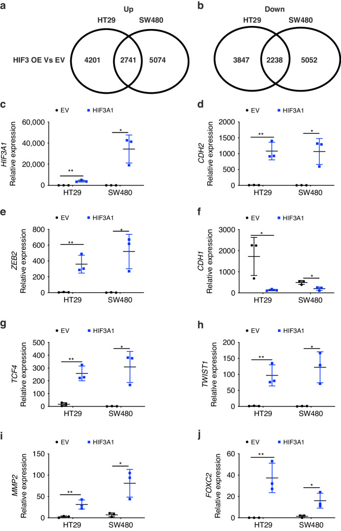
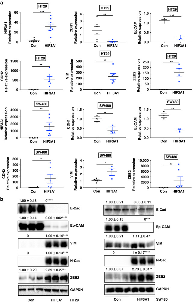
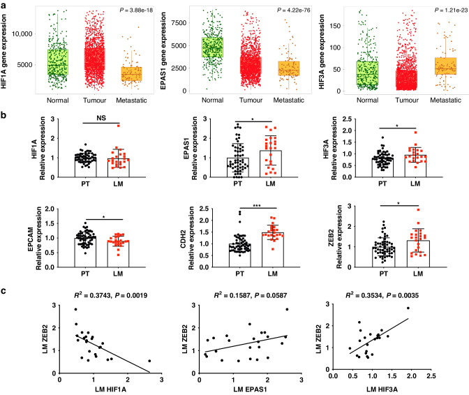
Background/objectives: Hypoxia-inducible factor (HIF)-3α1's role in colorectal cancer (CRC) cells, especially its effects on epithelial-mesenchymal transition (EMT), zinc finger E-box binding homeobox 2 (ZEB2) gene expression, and iron metabolism, remains largely unstudied. This research sought to elucidate these relationships.

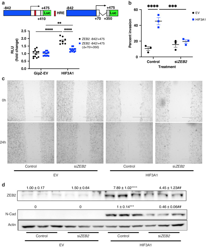
Methods: RNA-seq was conducted to investigate the impact of HIF-3α1 overexpression in CRC cells. Dual-luciferase reporter assays assessed the direct targeting of ZEB2 by HIF-3α1. Scratch assays measured changes in cell migration following HIF-3α1 overexpression and ZEB2 knockdown. The effects of HIF-3α1 overexpression on colon tumour growth and liver metastasis were examined in vivo. Iron chelation was used to explore the role of iron metabolism in HIF-3α1-mediated EMT and tumour growth.

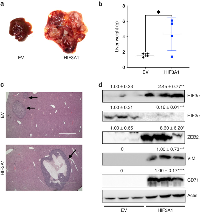
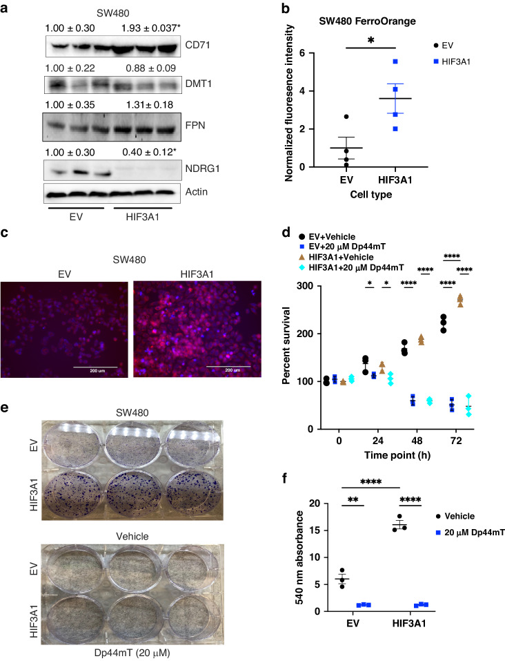
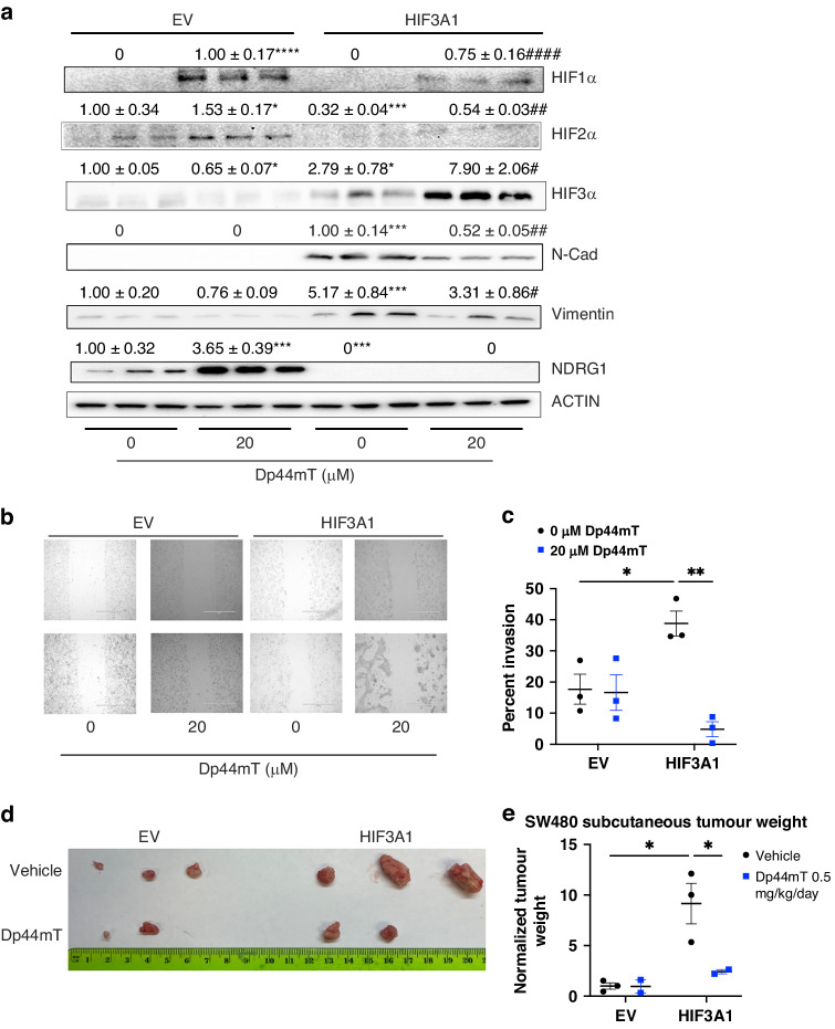
Results: HIF-3α1 overexpression induced EMT and upregulated ZEB2 expression, enhancing cancer cell migration. ZEB2 knockdown reduced mesenchymal markers and cell migration. HIF-3α1 promoted colon tumour growth and liver metastasis, increased transferrin receptor (TFRC) expression and cellular iron levels, and downregulated HIF-1α, HIF-2α, and NDRG1. Iron chelation mitigated HIF-3α1-mediated EMT, tumour growth, and survival.

Conclusions: HIF-3α1 plays a critical role in colon cancer progression by promoting EMT, iron accumulation, and metastasis through ZEB2 and TFRC regulation, suggesting potential therapeutic targets in CRC.


Conflict of interest statement

The authors declare no competing interests.

Figures

Fig. 1
Fig. 1. Overexpression of HIF3A1 in colon cancer cells promotes expression of EMT signature genes.
Venn Diagram for differential gene analysis of empty vector and HIF3A1 overexpressing HT29 and SW480 cells identifying commonly increased (a) and decreased (b) gene transcripts. cj Expression of HIF3A1, common EMT-associated genes (CDH2, ZEB2, TCF4, TWIST1, MMP2 and FOXC2) as well as epithelial marker CDH1 found after at least twofold change filtration and sorting by the most drastically changed. *P < 0.05 and **P < 0.01. Two-way ANOVA.
Fig. 2
Fig. 2. Validation of RNA-Seq results by qRT-PCR and western blot analysis.
a HIF3A1 overexpressing and control HT29 and SW480 colon cancer cells were analysed for expression of EMT signature genes by qRT-PCR. Results were calculated using the 2-ΔΔCt method using empty vector-transfected cells as reference. b Lysates prepared from HIF3A1 overexpressing and control HT29 (left) and SW480 (right) colon cancer cells were analysed for expression of EMT markers by western blot analysis. *P < 0.05, **P < 0.01, ***P < 0.001 and ****P < 0.0001. Student’s t test.
Fig. 3
Fig. 3. HIF3A is increased in CRC liver metastasis than primary tumour and is correlated with ZEB2 expression.
a Relative gene expression patterns of HIF1A, EPAS1, and HIF3A in normal, tumour, and metastatic samples. b Relative gene expression of HIF1A, EPAS1, HIF3A, EpCAM, CDH2 and ZEB2 in CRC patient samples of primary (PT) and liver metastasised (LM) tumours. c Correlation analysis relating gene expression levels of HIF1A, EPAS1 and HIF3A to ZEB2 in CRC patient LM samples. Gene expression was extracted from GSE28702. NS not significant, *P < 0.05 and ***P < 0.001. Student’s t test.
Fig. 4
Fig. 4. HIF3A1 induces ZEB2 transcription in an HRE-dependent manner.
Diagram of (a) the untruncated (−842/ + 475) and the truncated (Δ + 70/ + 350) ZEB2 5’-UTR luciferase reporter plasmid constructs and their corresponding luciferase assay results. The sequence corresponding to the ZEB2 promoter (−842/ + 1) is indicated in blue, the 5’-UTR intron (+ 410/ + 475) is indicated in grey, and the rest of the 5’-UTR is indicated in white. A putative HRE is indicated in red. Arrows indicate transcription start sites. Luc, luciferase. The relative position of the oligonucleotides used in this figure is indicated. b Graph of percent invasion of SW480 EV and HIF3A1 cells treated with control or esiZEB2. c Scratch assay and d western blotting analysis of SW480 cells transfected with control or esiZEB2 plasmid. **P < 0.01, ***P < 0.001, ****P < 0.0001. ##P < 0.01 compared to HIF3A1 control. Two-way ANOVA.
Fig. 5
Fig. 5. HIF3A1 increases CRC liver metastasis in mice.
a A representative gross liver image, b liver weights, c H&E staining of liver tissues and d western blotting analysis of liver tissues after intrasplenic injection of SW480 cells expressing EV or HIF3A1. Arrows indicate CRC liver metastatic sites. *P < 0.05, **P < 0.01, ***P < 0.001, ****P < 0.0001. Student’s t test.
Fig. 6
Fig. 6. HIF3A1 induces TFRC expression and increases iron content of CRC cells.
a Western blot analysis of iron metabolic proteins and metastasis suppressor NDRG1 in SW480 EV and HIF3A1 cells. b Quantification and c representative images of FerroOrange staining in SW480 EV and HIF3A1 cells. d MTT assay in SW480 EV and HIF3A1 cells treated with 0 (vehicle) or 20 μM Dp44mT. e Colony formation assay and f quantification in SW480 EV and HIF3A1 cells treated with 0 or 20 μM Dp44mT. *P < 0.05, **P < 0.01, and ****P < 0.0001. Student’s t test.
Fig. 7
Fig. 7. Dp44mT reduces mesenchymal markers, cell migration and tumour growth in HIF3A1 overexpressing cells.
a Western blotting analysis of SW480 EV or HIF3A1 cells treated with vehicle (DMSO) or Dp44mT at 20 μM. b Scratch assay and c quantification for SW480 cells treated with vehicle or Dp44mT (20 μM). d SW480 EV and HIF3A1 subcutaneous xenograft tumours images and e quantification from mice treated with 0.5 mg/kg/day of Dp44mT. *P < 0.05, **P < 0.01, ***P < 0.001 and ****P < 0.0001 compared to EV vehicle group or as indicated. #P < 0.05, ##P < 0.01, ####P < 0.0001 compared to HIF3A1 DMSO group. Two-way ANOVA.

Similar articles

Cited by

References

    1. Siegel RL, Miller KD, Jemal A. Cancer statistics, 2017. CA Cancer J Clin. 2017;67:7–30. doi: 10.3322/caac.21387. - DOI - PubMed
    1. Zarour LR, Anand S, Billingsley KG, Bisson WH, Cercek A, Clarke MF, et al. Colorectal cancer liver metastasis: evolving paradigms and future directions. Cell Mol Gastroenterol Hepatol. 2017;3:163–73. doi: 10.1016/j.jcmgh.2017.01.006. - DOI - PMC - PubMed
    1. Manfredi S, Lepage C, Hatem C, Coatmeyr O, Faivre J, Bouvier A. Epidemiology and management of liver metastases from colorectal cancer. Ann Surg. 2006;244:254–9. doi: 10.1097/01.sla.0000217629.94941.cf. - DOI - PMC - PubMed
    1. Leith JT, Padfield G, Faulkner L, Michelson S. Hypoxic fractions in xenografted human colon tumors. Cancer Res. 1991;51:5139–43. - PubMed
    1. Semenza GL. Hypoxia-inducible factors in physiology and medicine. Cell. 2012;148:399–408. doi: 10.1016/j.cell.2012.01.021. - DOI - PMC - PubMed

MeSH terms