. 2024 May 1;16(1):95.
doi: 10.1186/s13195-024-01462-3.

N, N-Dimethyltryptamine, a natural hallucinogen, ameliorates Alzheimer's disease by restoring neuronal Sigma-1 receptor-mediated endoplasmic reticulum-mitochondria crosstalk

Affiliations

N, N-Dimethyltryptamine, a natural hallucinogen, ameliorates Alzheimer's disease by restoring neuronal Sigma-1 receptor-mediated endoplasmic reticulum-mitochondria crosstalk

Dan Cheng et al. Alzheimers Res Ther. .

Abstract

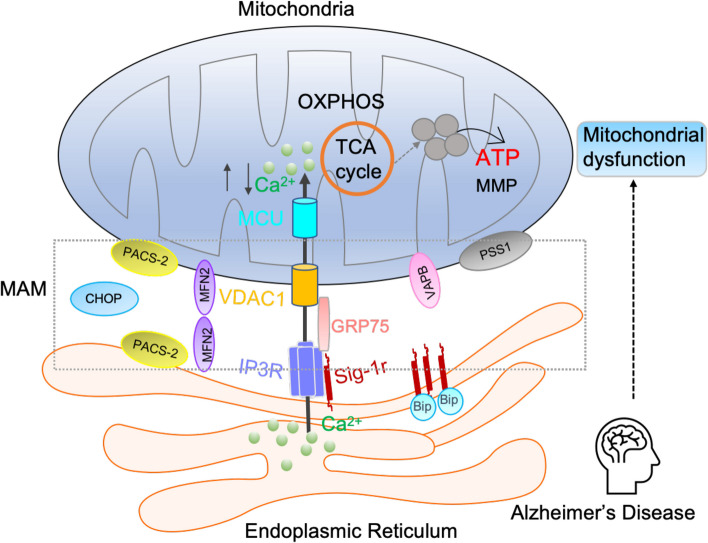
Background: Aberrant neuronal Sigma-1 receptor (Sig-1r)-mediated endoplasmic reticulum (ER)- mitochondria signaling plays a key role in the neuronal cytopathology of Alzheimer's disease (AD). The natural psychedelic N, N-dimethyltryptamine (DMT) is a Sig-1r agonist that may have the anti-AD potential through protecting neuronal ER-mitochondrial interplay.

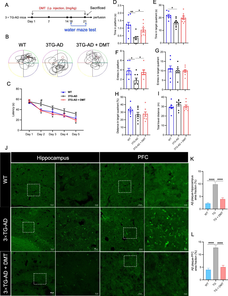
Methods: 3×TG-AD transgenic mice were administered with chronic DMT (2 mg/kg) for 3 weeks and then performed water maze test. The Aβ accumulation in the mice brain were determined. The Sig-1r level upon DMT treatment was tested. The effect of DMT on the ER-mitochondrial contacts site and multiple mitochondria-associated membrane (MAM)-associated proteins were examined. The effect of DMT on calcium transport between ER and mitochondria and the mitochondrial function were also evaluated.

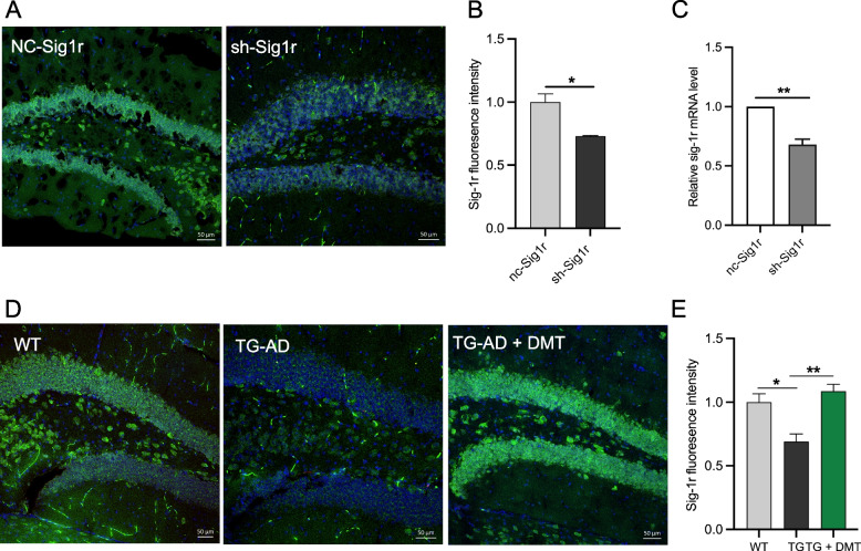
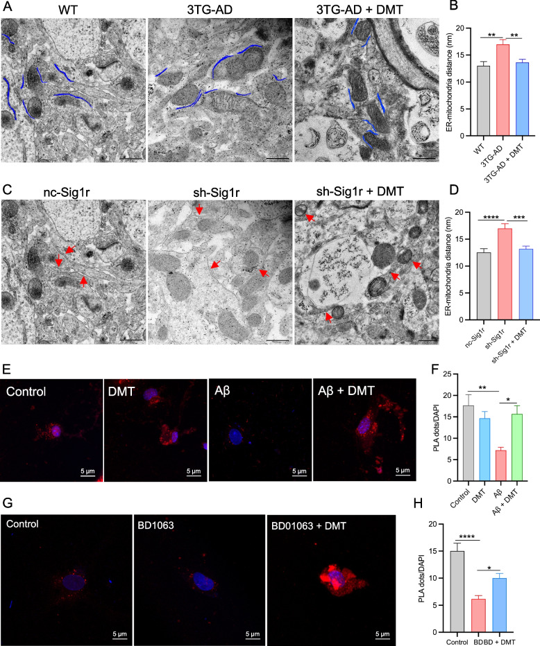
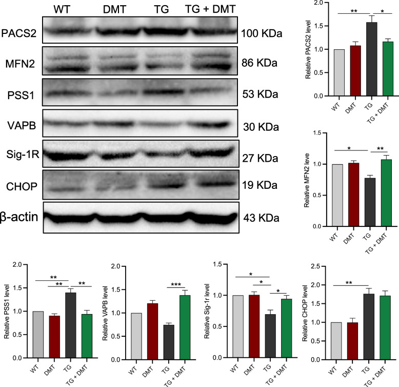
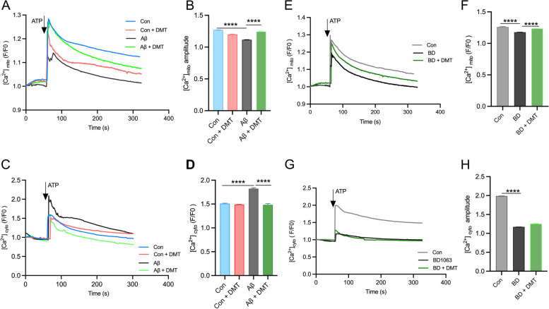
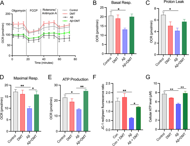
Results: chronic DMT (2 mg/kg) markedly alleviated cognitive impairment of 3×TG-AD mice. In parallel, it largely diminished Aβ accumulation in the hippocampus and prefrontal cortex. DMT restored the decreased Sig-1r levels of 3×TG-AD transgenic mice. The hallucinogen reinstated the expression of multiple MAM-associated proteins in the brain of 3×TG-AD mice. DMT also prevented physical contact and calcium dynamic between the two organelles in in vitro and in vivo pathological circumstances. DMT modulated oxidative phosphorylation (OXPHOS) and ATP synthase in the in vitro model of AD.

Conclusion: The anti-AD effects of DMT are associated with its protection of neuronal ER-mitochondria crosstalk via the activation of Sig-1r. DMT has the potential to serve as a novel preventive and therapeutic agent against AD.

Keywords: Alzheimer’s disease; ER-mitochondria crosstalk; N,N-Dimethyltryptamine; Sigma-1 receptor, calcium homeostasis; cognitive impairment.


Conflict of interest statement

The authors declare no competing interests.

Figures

Fig. 1
Fig. 1
DMT improves cognitive impairment and diminishes Aβ pathology in the 3×TG-AD mice. A Experimental timeline. B The representative swim paths of mice. C Latency to platform of mice during the training trial. D Time in platform, E Time in target quadrant, F Entries in platform, G Entries in target quadrant, H Distance in target quadrant, I Total travel distance of mice in the probe test. (n = 8 in each group). J The representative images of Aβ plaque in the hippocampus and prefrontal cortex (PFC) of 3×TG-AD mice. The images on the right are enlarged views of the image on the left in each panel. Scale bar: 50 μm (left), 10 μm (right). K, L The quantification of the Aβ accumulation in the hippocampus and PFC, respectively. The results were presented as the mean area fraction of the positive particle. In the training trials of water maze test, data were analyzed using two-way ANOVA test; in the probe test, data were analyzed using one-way ANOVA. Data analysis of Aβ plaque was performed on average 5 randomly selected sections from each mouse (n = 3 per group) using one-way ANOVA. *P < 0.05, **P < 0.01, ****P < 0.0001. TG, 3×TG-AD
Fig. 2
Fig. 2
DMT improves the Sig-1r levels in the mice hippocampus and primary hippocampal neurons. A, B Representatives and the quantification of the Sig-1r level in the hippocampus of sh-Sig1r knockdown mice. C The mRNA level of Sig-1r in the hippocampus of sh-Sig1r knockdown mice. D, E Representative images and the quantification of the Sig-1r level in the hippocampus of 3×TG-AD mice. In the in vivo study, data was obtained from average 5 randomly selected sections from each mouse (n = 3 per group). Data was analyzed using one-way ANOVA. *P < 0.05, **P < 0.01. TG, 3×TG-AD
Fig. 3
Fig. 3
DMT modulates endoplasmic reticulum (ER) – mitochondrial distance and contacts. Two different animal models, 3×TG-AD mice and sh-Sig1r knockdown mice were used. A, B Representatives and quantification of ER-mitochondrial distance in the hippocampus of 3×TG-AD mice. C, D Representatives and quantitative analysis of ER-mitochondrial distance in the hippocampus of sh-Sig1r knock down mice. Scale bar: 500 nm. Data was obtained from 3 mice per group with 10 images in each mouse. E, F Representatives and quantification of the PLA signal in the cells exposed to Aβ with or without DMT treatment. G, H Representatives and quantification of the PLA signal in the cells exposed to BD1063 with or without DMT treatment. Scale bar: 5μm. Data was representative of 3 independent experiments and analyzed using one-way ANOVA. *P < 0.05, **P < 0.01, ***P < 0.001, ****P < 0.0001
Fig. 4
Fig. 4
DMT modulates the expression of MAM-associated proteins in 3×TG-AD mice brains. Western blot was used to determine the expression of MAM-associated proteins. Data were analyzed using one-way ANOVA (n = 3 per group). *P < 0.05, **P < 0.01, ***P < 0.001
Fig. 5
Fig. 5
DMT modulates the calcium transfer between ER and mitochondria. Cytosolic and mitochondrial Ca2+ were measured in the primary hippocampus by using live cell calcium imaging. A, B and C, D show the mitochondrial and cytosolic calcium, respectively, in the cells exposed to Aβ with or without DMT treatment. E, F and G, H show the mitochondrial and cytosolic calcium, respectively, in the cells exposed to BD1063 with or without DMT treatment. A, C, E, G show the representative figure of Ca2+ peaks after ATP treatment. B, D, F, H show the the amplitude peak of calcium levels after ATP treatment. Data were obtained from 3 independent experiments and analyzed using two-way ANOVA. [Ca2+]mito, mitochondrial calcium; [Ca2+]cyto, cytosolic calcium. BD, BD1063. ****P < 0.0001
Fig. 6
Fig. 6
DMT rescues mitochondrial dysfunction in the primary hippocampal neurons. A Oxygen consumption rate (OCR) at baseline and following: oligomycin, FCCP, and rotenone + antimycin A. B, C, D, E show the quantification of basal respiration, proton leak, maximal respiration, and ATP production. F shows the quantification of the mitochondrial membrane potential (MMP). G shows the quantification of the cellular ATP level. Data were obtained from 3 independent experiments and analyzed using two-way ANOVA. *P < 0.05, **P < 0.01
Fig. 7
Fig. 7
The putative mechanism of DMT improving the spatial learning and memory in animal models of AD. The current study demonstrated that the anti-AD effects of DMT are associated with its restoration of neuronal ER-mitochondria crosstalk via the Sig-1r activation. DMT modulates the mitochondrial calcium uptake and ER-mitochondrial contacts in the in vivo and in vitro models, facilitates the TCA cycle, and protects against mitochondrial dysfunction in Alzheimer’ disease

References

    1. Galvin JE, et al. The social and economic burden of frontotemporal degeneration. Neurol. 2017;89(20):2049–2056. - PMC - PubMed
    1. Athar T, Al Balushi K, Khan SA. Recent advances on drug development and emerging therapeutic agents for Alzheimer’s disease. Molecular biol rep. 2021;48(7):5629–5645. - PMC - PubMed
    1. Eysert F, et al. Molecular dysfunctions of mitochondria-associated membranes (MAMs) in Alzheimer’s disease. Int J Molecular Sci. 2020;21(24):9521. - PMC - PubMed
    1. Markovinovic A, et al. Endoplasmic reticulum–mitochondria signaling in neurons and neurodegenerative diseases. J Cell Sci. 2022;135(3):pjcs248534. - PubMed
    1. Hedskog L, et al. Modulation of the endoplasmic reticulum–mitochondria interface in Alzheimer’s disease and related models. Proceed Nat Academ Sci. 2013;110(19):7916–7921. - PMC - PubMed

Publication types