Review
. 2024 May 1;22(1):249.
doi: 10.1186/s12964-024-01625-7.

Cuproptosis: unveiling a new frontier in cancer biology and therapeutics

Affiliations
Review

Cuproptosis: unveiling a new frontier in cancer biology and therapeutics

Ying Feng et al. Cell Commun Signal. .

Abstract

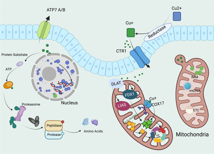
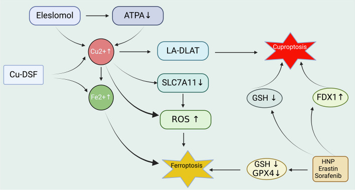
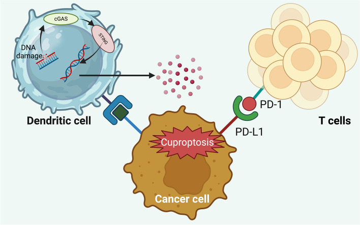
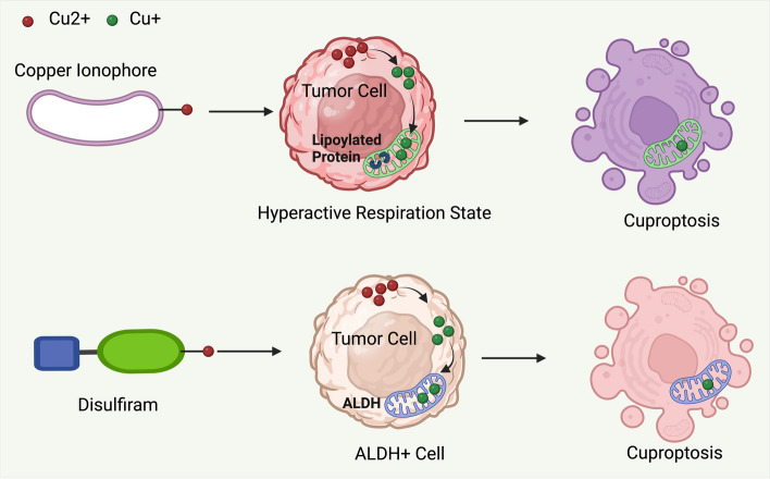
Copper plays vital roles in numerous cellular processes and its imbalance can lead to oxidative stress and dysfunction. Recent research has unveiled a unique form of copper-induced cell death, termed cuproptosis, which differs from known cell death mechanisms. This process involves the interaction of copper with lipoylated tricarboxylic acid cycle enzymes, causing protein aggregation and cell death. Recently, a growing number of studies have explored the link between cuproptosis and cancer development. This review comprehensively examines the systemic and cellular metabolism of copper, including tumor-related signaling pathways influenced by copper. It delves into the discovery and mechanisms of cuproptosis and its connection to various cancers. Additionally, the review suggests potential cancer treatments using copper ionophores that induce cuproptosis, in combination with small molecule drugs, for precision therapy in specific cancer types.

Keywords: Copper; Copper homeostasis; Cuproptosis; Immunotherapy; Mitochondria; Targetted therapy.


Conflict of interest statement

The authors declare no competing interests.

Figures

Fig. 1
Fig. 1
The timeline of the discovery of cuproptosis. Abbreviation: ROS, reactive oxygen species; DSF, Disulfiram; FDX1, Ferredoxin 1; LA, Lipoic Acid
Fig. 2
Fig. 2
Copper metabolism and homeostasis in the human body. The intricate mechanism of copper absorption and metabolism within the human body commences with the ingestion of dietary Cu(II), which is absorbed in the stomach and small intestine. Subsequent to absorption, Cu(II) undergoes reduction to Cu(I) by a reductase enzyme. This monovalent copper is then bound by chaperone proteins, notably ATOX1, and is transported to the liver for further utilization and regulation. Within the liver, ATP7B plays an instrumental role in the integration of copper into ceruloplasmin, which serves as a carrier for copper in the bloodstream to various tissues, and in mediating the excretion of surplus copper into bile for elimination. The function of liver is crucial in preserving copper homeostasis, ensuring the prevention of both copper deficiency and toxicity. Additionally, another transporter, ATP7A, is primarily tasked with exporting copper from intestinal epithelial cells into the bloodstream, aiding in the oxidation of Cu(I) back to Cu(II), which facilitates its release and distribution throughout the body. Abbreviation: ATOX1, Antioxidant 1 Copper Chaperone
Fig. 3
Fig. 3
Intracellular copper distribution pathways. The intracellular copper metabolism signaling pathway involves a sequence of events starting with the reduction of extracellular Cu(II) to Cu(I), which is then transported into the cell via the CTR1. Once inside, Cu(I) associates with the copper chaperone for CCS and SOD1, facilitating its distribution to various subcellular compartments. Additionally, Cu(I) interacts with ATOX1, predominantly directing it to ATP7A and ATP7B, for transport to the Golgi apparatus. A minor portion of Cu(I) binds to transcription factors in the nucleus, influencing gene expression. Cu(I) also binds to COX17 and is transported to CCO in mitochondria, contributing to the oxidative phosphorylation process. Surplus Cu(I) is expelled from the cell through ATP7A/B on the plasma membrane
Fig. 4
Fig. 4
Molecular mechanisms of cuproptosis. The molecular mechanisms underlying cuproptosis involve several key processes facilitated by copper ions. i. Cu(II) outside the cell are reverted to Cu(I) by a reductase enzyme. This oxidization-reduction form of copper is then transported into the cell by CTR1. ii. Copper is handled by various chaperone proteins to prevent toxic accumulation inside cell. ATP7A/B can pump excess copper out of the cell or deliver it to the Golgi apparatus for incorporation into copper-dependent enzymes. iii. Copper plays an essential role in several biochemical pathways inside the mitochondria. Here, it is involved in the ETC, which is a series of protein complexes embedded in the inner mitochondrial membrane. iv. The copper chaperone COX17 delivers copper to the mitochondrial enzyme complexes, such as COO for their proper function. The mitochondrion matrix contains several enzymes, including LIAS and DLAT, which are part of the TCA cycle. v. FDX1 is involved in the transfer of electrons in metabolic processes and is believed to interact with copper ions directly or indirectly affecting the TCA cycle. vi.the presence of mitochondrial DNA suggests that changes in copper homeostasis might impact mitochondrial genetics and, consequently, the function of organelle. Abbreviation: ETC, electron transport chain; IMM, inner mitochondrial membrane; mtDNA, mitochondrial DNA; LIAS, Lipoic Acid Synthase;DLAT; Dihydrolipoamide S-Acetyltransferase
Fig. 5
Fig. 5
Timeline of key discoveries related to cell death. The exploration of cell death mechanisms has unveiled numerous modes beyond traditional necrosis, each with distinct triggers and cellular responses. Apoptosis, identified in the 1970s, emerged as the first form of programmed cell death, pivotal for development and disease. The 2000s introduced autophagy and necroptosis, highlighting the complexity of cell death and its role in health and pathology. Discoveries like cuproptosis in recent years have further expanded our understanding, revealing the intricate balance cells maintain between survival and death. These advancements underscore the diverse cellular strategies for self-destruction, critical for therapeutic targeting in diseases. Abbreviation: LDCD, lysosome-dependent cell death; NETosis, neutrophil extracellular traps; ICD, immunogenic cell death
Fig. 6
Fig. 6
The crosstalk between ferroptosis and cuproptosis inside mitochondria. TCA cycle within mitochondria acts as a co-regulator for both cystine-depleted ferroptosis and cuproptosis. The enzymes COX7A1 and FAK are shown to amplify the mitochondrial TCA cycle, thereby enhancing production and promoting the onset of both cystine-depleted ferroptosis and cuproptosis. Cuproptosis is depicted as dependent on mitochondrial respiratory activities and is shown to be inhibited by specific mitochondrial respiration inhibitors such as rotenone, antimycin, and UK5099. Abbreviation: COX7A1, cytochrome c oxidase subunit 7A1; FAK, focal adhesion kinase; DLAT, dihydrolipoamide s-acetyltransferase; etc., electron transport chain. Created with BioRender
Fig. 7
Fig. 7
The crosstalk between ferroptosis and cuproptosis mechanisms in cancer. ES increases intracellular Cu(II) concentration and inhibits ATP7A, thus contributing to cuproptosis. Concurrently, Cu-DSF complex elevates Cu(II) and Fe(II) levels, which would lead to an increase in ROS. Increased Cu (II) impairs LA-DLAT, which is linked to cuproptosis, while decreased expression of SLC7A11 reduces GSH levels. This increase in ROS, stemming from both the elevation of Fe (II) and the decrease in SLC7A11, would trigger ferroptosis, which is a form of cell death characterized by lipid peroxidation due to iron accumulation. A decrease in GSH and GPX4 also further drives the cell towards ferroptosis, while levated FDX1 levels facilitate cuproptosis. Abbreviation:ATPA, adenosine triphosphatase; HNP, hollow nano platform. Created with BioRender
Fig. 8
Fig. 8
Cuproptosis enhances anti-cancer immune effects by influencing the cGAS-STING pathway. This signaling pathway is activated inside dendritic cells by cancer cells undergoing cuproptosis, triggered by treatments such as ES and copper chloride (CuCl2). This reaction leads to the secretion of pro-inflammatory cytokines and chemokines including IL-2, TNF-α (tumor necrosis factor-alpha), IFN-γ (interferon-gamma), C-X-C motif chemokine ligand (CXCL) 10, and CXCL11. Furthermore, the concurrent use of cuproptosis inducers with PD-1 blockade significantly increases the population of circulating CD45 + CD8 + T cells, thereby enhancing the overall effectiveness of cancer therapy
Fig. 9
Fig. 9
Two potential therapeutic approaches aim to harness cuproptosis for cancer treatment. Firstly, the copper ionophore ES is posited to trigger cuproptosis in cancer cells characterized by elevated expression of lipoylated mitochondrial enzymes or those in an enhanced respiratory state. Secondly, the combination of DSF and copper specifically targets cancer cells exhibiting high levels of ALDH expression, presenting a targeted strategy against cancer cells with this specific metabolic profile

Similar articles

Cited by

References

    1. Tsvetkov P, Coy S, Petrova B, Dreishpoon M, Verma A, Abdusamad M, et al. Copper induces cell death by targeting lipoylated TCA cycle proteins. Science. 2022;375:1254–61. doi: 10.1126/science.abf0529. - DOI - PMC - PubMed
    1. Helman SL, Zhou J, Fuqua BK, Lu Y, Collins JF, Chen H, et al. The biology of mammalian multi-copper ferroxidases. Biometals. 2023;36:263–81. doi: 10.1007/s10534-022-00370-z. - DOI - PMC - PubMed
    1. Xue Q, Kang R, Klionsky DJ, Tang D, Liu J, Chen X. Copper metabolism in cell death and autophagy. Autophagy. 2023;19:2175–95. doi: 10.1080/15548627.2023.2200554. - DOI - PMC - PubMed
    1. Yang Y, Li M, Chen G, Liu S, Guo H, Dong X, et al. Dissecting copper biology and cancer treatment: ‘Activating Cuproptosis or suppressing Cuproplasia’. Coord Chem Rev. 2023;495:215395. doi: 10.1016/j.ccr.2023.215395. - DOI
    1. Zugazagoitia J, Guedes C, Ponce S, Ferrer I, Molina-Pinelo S, Paz-Ares L. Current Challenges in Cancer Treatment. Clin Ther. 2016;38:1551–66. doi: 10.1016/j.clinthera.2016.03.026. - DOI - PubMed

Publication types