Review
. 2024 May 1;22(1):215.
doi: 10.1186/s12951-024-02455-y.

Advances in the application of extracellular vesicles derived from three-dimensional culture of stem cells

Affiliations
Review

Advances in the application of extracellular vesicles derived from three-dimensional culture of stem cells

Wenya Chen et al. J Nanobiotechnology. .

Abstract

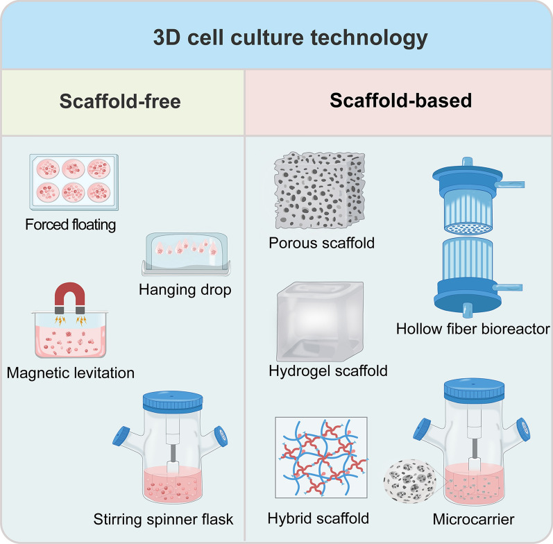
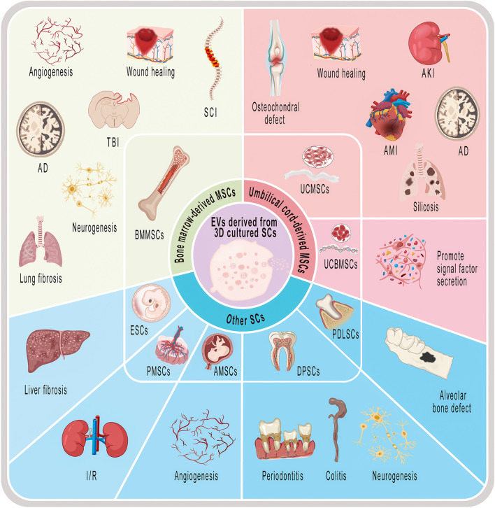
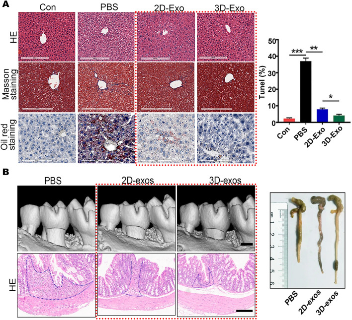
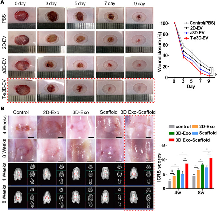
Stem cells (SCs) have been used therapeutically for decades, yet their applications are limited by factors such as the risk of immune rejection and potential tumorigenicity. Extracellular vesicles (EVs), a key paracrine component of stem cell potency, overcome the drawbacks of stem cell applications as a cell-free therapeutic agent and play an important role in treating various diseases. However, EVs derived from two-dimensional (2D) planar culture of SCs have low yield and face challenges in large-scale production, which hinders the clinical translation of EVs. Three-dimensional (3D) culture, given its ability to more realistically simulate the in vivo environment, can not only expand SCs in large quantities, but also improve the yield and activity of EVs, changing the content of EVs and improving their therapeutic effects. In this review, we briefly describe the advantages of EVs and EV-related clinical applications, provide an overview of 3D cell culture, and finally focus on specific applications and future perspectives of EVs derived from 3D culture of different SCs.

Keywords: 3D cell culture; Clinical applications; Extracellular vesicles; Stem cells; Therapeutics.


Conflict of interest statement

The authors declare that they have no competing interests.

Figures

Fig. 1
Fig. 1
Available 3D cell culture technologies. They are mainly divided into scaffold-free and scaffold-based technologies. Scaffold-free technologies include forced floating (ultra-low attachment plate), hanging drop, magnetic levitation and stirring spinner flask. Scaffold-based technologies include porous and hydrogel scaffolds made from natural or synthetic materials, hybrid scaffolds, hollow fiber bioreactors and microcarrier-based bioreactors
Fig. 2
Fig. 2
Current applications of EVs derived from 3D cultured SCs. EVs derived from 3D culture of BMMSCs for wound healing, angiogenesis, neurogenesis, SCI, TBI, AD, lung fibrosis; EVs derived from 3D culture of UCMSCs for osteochondral defect, wound healing, AKI, AD, AMI and silicosis; EVs derived from 3D culture of UCBMSCs for promoting signal factor secretion. EVs derived from 3D culture of ESCs for liver fibrosis; EVs derived from 3D culture of PMSCs for I/R; EVs derived from 3D culture of AMSCs for angiogenesis; EVs derived from 3D culture of DPSCs for periodontitis, colitis and neurogenesis; EVs derived from 3D culture of PDLSCs for alveolar bone defect
Fig. 3
Fig. 3
Applications of EVs derived from 3D cultured BMMSCs. A SD rat BMMSC-derived 3D-Exos applied to SCI rat model. Representative HE staining of tissue damage following SCI insult. Quantitative analysis of cavity volume in each group. Reproduced with permission from reference [43]. Copyright 2022, American Chemical Society B HBMMSC-derived 3D-Exos applied to TBI rat model. CD68 and GFAP staining and statistics on the number of positive cells in rat brain tissues. Reproduced with permission from reference [161]. Copyright 2017, Elsevier
Fig. 4
Fig. 4
Applications of EVs derived from 3D cultured UCMSCs. A UCMSC-derived 3D-Exos applied to rabbit cartilage defect model. Representative macroscopic images of the regenerated tissues. Staining results of HE, TB, Saf-O and immunohistochemical staining for type II collagens. Wakitani scores for the histological sections. Reproduced with permission from reference [142]. Copyright 2020, Springer Nature B HucMSC-derived 3D-Exos applied to AKI mouse model. Representative images of PAS staining of renal cortex. Representative immunostaining images of CD68+ macrophages or CD3+ T cells in the tubulointerstitium. Serum creatinine graph. Reproduced with permission from reference [28]. Copyright 2020, Springer Nature
Fig. 5
Fig. 5
Applications of EVs derived from 3D cultured hESCs and hDPSCs. A HESC-derived 3D-Exos applied to liver fibrosis mice model. Representative images of HE, masson and oil red staining of liver tissue. Quantification of positive cells in TUNEL staining of liver tissues. Reproduced with permission from reference [42]. Copyright 2021, Springer Nature B HDPSC-derived 3D-Exos applied to periodontitis and colitis model. 3D reconstructions of maxillae revealed by micro-CT. HE staining showed histopathological changes in the colon. Representative colon pictures of the mice in each group. Reproduced with permission from reference [44]. Copyright 2021, Springer Nature
Fig. 6
Fig. 6
Methods to improve the efficacy of 3D-EVs. A HucMSC-derived T-a3D-EVs applied to wound healing mouse model. Representative in vivo wound closing images. Quantitative wound closure rate. Reproduced with permission from reference [147]. Copyright 2023, Elsevier B HucMSC-derived 3D-Exos combined scaffolds applied to rat knee osteochondral defect model. Microscopic observation of the repaired tissues. Micro-CT images showing 2D and 3D reconstructions of the repaired cartilage. ICRS score of the cartilage defect. Reproduced with permission from reference [164]. Copyright 2023, Elsevier

Similar articles

Cited by

References

    1. Sebastiao MJ, Serra M, Gomes-Alves P, Alves PM. Stem cells characterization: OMICS reinforcing analytics. Curr Opin Biotechnol. 2021;71:175–181. doi: 10.1016/j.copbio.2021.07.021. - DOI - PubMed
    1. Zhou C, Zhang B, Yang Y, Jiang Q, Li T, Gong J, et al. Stem cell-derived exosomes: emerging therapeutic opportunities for wound healing. Stem Cell Res Ther. 2023;14:107. doi: 10.1186/s13287-023-03345-0. - DOI - PMC - PubMed
    1. Zakrzewski W, Dobrzynski M, Szymonowicz M, Rybak Z. Stem cells: past, present, and future. Stem Cell Res Ther. 2019;10:68. doi: 10.1186/s13287-019-1165-5. - DOI - PMC - PubMed
    1. Kimbrel EA, Lanza R. Next-generation stem cells—ushering in a new era of cell-based therapies. Nat Rev Drug Discov. 2020;19:463–479. doi: 10.1038/s41573-020-0064-x. - DOI - PubMed
    1. Hu C, Zhao L, Zhang L, Bao Q, Li L. Mesenchymal stem cell-based cell-free strategies: safe and effective treatments for liver injury. Stem Cell Res Ther. 2020;11:377. doi: 10.1186/s13287-020-01895-1. - DOI - PMC - PubMed

MeSH terms