

Bone marrow mesenchymal stem cell-derived exosomes shuttle microRNAs to endometrial stromal fibroblasts that promote tissue proliferation /regeneration/ and inhibit differentiation
- PMID: 38693588
- PMCID: PMC11064399
- DOI: 10.1186/s13287-024-03716-1
Bone marrow mesenchymal stem cell-derived exosomes shuttle microRNAs to endometrial stromal fibroblasts that promote tissue proliferation /regeneration/ and inhibit differentiation
Abstract
Background: Human bone marrow-derived stem cells (hBMDSCs) are well characterized mediators of tissue repair and regeneration. An increasing body of evidence indicates that these cells exert their therapeutic effects largely through their paracrine actions rather than clonal expansion and differentiation. Here we studied the role of microRNAs (miRNAs) present in extracellular vesicles (EVs) from hBMDSCs in tissue regeneration and cell differentiation targeting endometrial stromal fibroblasts (eSF).
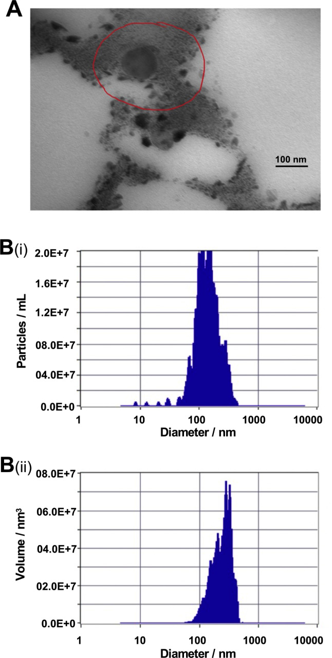
Methods: Extracellular vesicles (EVs) are isolated from hBMDSCs, characterized by transmission electron microscopy (TEM) and nanoparticle tracking analysis (NTA) techniques. Extracted total RNA from EVs was subjected to RNA seq analysis. Transfection and decidualization studies were carried out in endometrial stromal fibroblasts (eSF). Gene expression was analyzed by qRTPCR. Unpaired t-test with Welch's correction was used for data analysis between two groups.
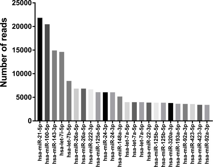
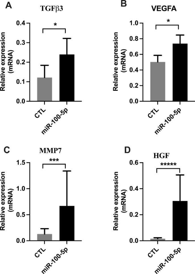
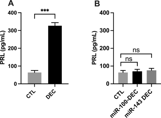
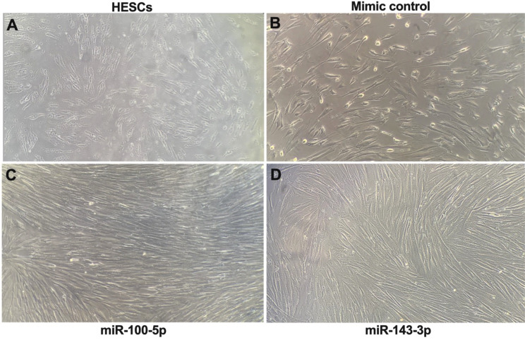
Results: We identified several microRNAs (miRNAs) that were highly expressed, including miR-21-5p, miR-100-5p, miR-143-3p and let7. MiR-21 is associated with several signaling pathways involved in tissue regeneration, quiescence, cellular senescence, and fibrosis. Both miR-100-5p and miR-143-3p promoted cell proliferation. MiR-100-5p specifically promoted regenerative processes by upregulating TGF-ß3, VEGFA, MMP7, and HGF. MiR-100-5p blocked differentiation or decidualization as evidenced by morphologic changes and downregulation of decidualization mediators including HOXA10, IGFBP1, PRL, PR-B, and PR.
Conclusion: EVs delivered to tissues by hBMDSCs contain specific miRNAs that prevent terminal differentiation and drive repair and regeneration. Delivery of microRNAs is a novel treatment paradigm with the potential to replace BMDSCs in cell-free regenerative therapies.
Keywords: BMDSCs; Decidualization; Exosomes; eSF; miRNAs.
© 2024. The Author(s).
Conflict of interest statement
All authors declare no competing interests.
Figures


Similar articles
-
miR-340-3p-modified bone marrow mesenchymal stem cell-derived exosomes inhibit ferroptosis through METTL3-mediated m6A modification of HMOX1 to promote recovery of injured rat uterus.Stem Cell Res Ther. 2024 Jul 29;15(1):224. doi: 10.1186/s13287-024-03846-6. Stem Cell Res Ther. 2024. PMID: 39075530 Free PMC article.
-
Isolation and characterization of bone mesenchymal cell small extracellular vesicles using a novel mouse model.J Bone Miner Res. 2024 Oct 29;39(11):1633-1643. doi: 10.1093/jbmr/zjae135. J Bone Miner Res. 2024. PMID: 39173022 Free PMC article.
-
miR-144-3p targeting FLRT3 in osteogenic differentiation of mandibular bone marrow mesenchymal stem cells.Hum Genomics. 2025 Jul 13;19(1):79. doi: 10.1186/s40246-025-00788-9. Hum Genomics. 2025. PMID: 40653464 Free PMC article.
-
MicroRNAs in oral fluids (saliva and gingival crevicular fluid) as biomarkers in orthodontics: systematic review and integrated bioinformatic analysis.Prog Orthod. 2021 Oct 11;22(1):31. doi: 10.1186/s40510-021-00377-1. Prog Orthod. 2021. PMID: 34632546 Free PMC article.
-
Expression and Regulatory Mechanisms of MicroRNA in Cholesteatoma: A Systematic Review.Int J Mol Sci. 2023 Jul 31;24(15):12277. doi: 10.3390/ijms241512277. Int J Mol Sci. 2023. PMID: 37569652 Free PMC article.
Cited by
-
Application of biomaterials in mesenchymal stem cell based endometrial reconstruction: current status and challenges.Front Bioeng Biotechnol. 2025 Jan 29;13:1518398. doi: 10.3389/fbioe.2025.1518398. eCollection 2025. Front Bioeng Biotechnol. 2025. PMID: 39944223 Free PMC article. Review.
-
sEV-mediated lipid droplets transferred from bone marrow adipocytes promote ferroptosis and impair osteoblast function.J Lipid Res. 2024 Nov;65(11):100657. doi: 10.1016/j.jlr.2024.100657. Epub 2024 Sep 24. J Lipid Res. 2024. PMID: 39326787 Free PMC article.
References
Publication types
MeSH terms
Substances
Grants and funding
LinkOut - more resources
Full Text Sources
Research Materials
