

Correlation of age with the size of subcortical nuclei of the brain and its implication in degenerative disease: A magnetic resonance imaging study
- PMID: 38693963
- PMCID: PMC11061590
- DOI: 10.12688/f1000research.139515.2
Correlation of age with the size of subcortical nuclei of the brain and its implication in degenerative disease: A magnetic resonance imaging study
Abstract
Background: Aging is a non-modifiable risk factor for neurodegenerative disease. It is well established that the brain undergoes physiological atrophy with age. So, this study was conducted to analyse the correlation between the age of the person and the size of the various subcortical nuclei of the brain and whether these measurements can serve as a useful indicator for physiological atrophy leading to degenerative disease in clinical practice.
Methods: A total of 600 MRI scans from healthy individuals were examined and the measurements of subcortical nuclei were taken and subsequently analysed.
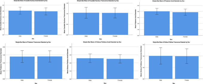
Results: A statistically significant difference between the genders was observed in the sizes of the axial diameters of caudate nucleus, putamen and globus pallidus. Caudate nucleus transverse diameter showed a moderate negative correlation with age in males. Globus pallidus axial diameter with age showed weak positive correlation for males. Globus pallidus transverse diameter showed weak positive correlation with age for both males and females, but it was stronger for males compared to females.
Conclusions: These results will help neurologists and neurosurgeons in analysing various early degenerative diseases and treat them accordingly.
Keywords: Aging; Brain; Magnetic resonance imaging; Neurodegenerative diseases; Neurosurgeons..
Copyright: © 2024 Dhamija A et al.
Conflict of interest statement
No competing interests were disclosed.
Figures


Similar articles
-
Developmentally stable whole-brain volume reductions and developmentally sensitive caudate and putamen volume alterations in those with attention-deficit/hyperactivity disorder and their unaffected siblings.JAMA Psychiatry. 2015 May;72(5):490-9. doi: 10.1001/jamapsychiatry.2014.3162. JAMA Psychiatry. 2015. PMID: 25785435
-
Cortical pencil lining on SWI MRI in NBIA and healthy aging.BMC Neurol. 2019 Oct 14;19(1):233. doi: 10.1186/s12883-019-1471-7. BMC Neurol. 2019. PMID: 31607263 Free PMC article.
-
Caudate nucleus hypointensity in the elderly is associated with markers of neurodegeneration on MRI.Neurobiol Aging. 2008 Dec;29(12):1839-46. doi: 10.1016/j.neurobiolaging.2007.05.008. Epub 2007 Jun 28. Neurobiol Aging. 2008. PMID: 17599695
-
Regional Cerebral Gray Matter Volume in HIV-Positive Patients with Executive Function Deficits.J Neuroimaging. 2016 Jul;26(4):450-7. doi: 10.1111/jon.12327. Epub 2016 Jan 19. J Neuroimaging. 2016. PMID: 26780881
-
Age, gender, and hemispheric differences in human striatum: a quantitative review and new data from in vivo MRI morphometry.Neurobiol Learn Mem. 1995 Mar;63(2):133-42. doi: 10.1006/nlme.1995.1013. Neurobiol Learn Mem. 1995. PMID: 7663886 Review.
References
-
- Almeida OP, Burton EJ, McKeith I, et al. : MRI study of caudate nucleus volume in Parkinson’s disease with and without dementia with Lewy bodies and Alzheimer’s disease. Dement. Geriatr. Cogn. Disord. 2003;16(2):57–63. - PubMed
-
- Geng D-Y, Li Y-X, Zee C-S: Magnetic resonance imaging-based volumetric analysis of basal ganglia nuclei and substantia nigra in patients with Parkinson’s disease. Neurosurgery. 2006;58(2):256–262. - PubMed
-
- Lisanby SH, McDonald WM, Massey EW, et al. : Diminished subcortical nuclei volumes in Parkinson’s disease by MR imaging. J. Neural. Transm. Suppl. 1993;40:13–21. - PubMed
Publication types
MeSH terms
Associated data
LinkOut - more resources
Full Text Sources
Medical