. 2024 Apr 17:18:1363219.
doi: 10.3389/fncel.2024.1363219. eCollection 2024.

Cyclic AMP signaling promotes regeneration of cochlear synapses after excitotoxic or noise trauma

Affiliations

Cyclic AMP signaling promotes regeneration of cochlear synapses after excitotoxic or noise trauma

Sriram Hemachandran et al. Front Cell Neurosci. .

Abstract

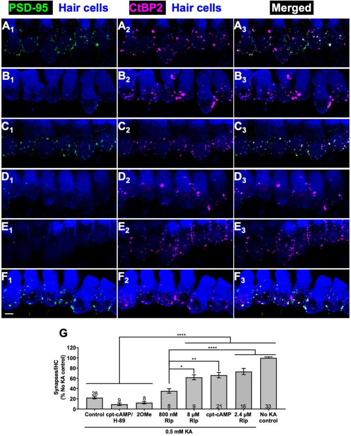
Introduction: Cochlear afferent synapses connecting inner hair cells to spiral ganglion neurons are susceptible to excitotoxic trauma on exposure to loud sound, resulting in a noise-induced cochlear synaptopathy (NICS). Here we assessed the ability of cyclic AMP-dependent protein kinase (PKA) signaling to promote cochlear synapse regeneration, inferred from its ability to promote axon regeneration in axotomized CNS neurons, another system refractory to regeneration.

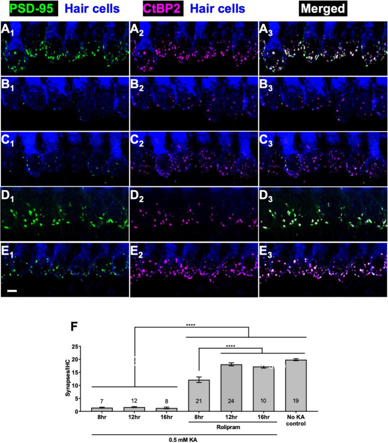
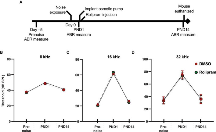
Methods: We mimicked NICS in vitro by applying a glutamate receptor agonist, kainic acid (KA) to organotypic cochlear explant cultures and experimentally manipulated cAMP signaling to determine whether PKA could promote synapse regeneration. We then delivered the cAMP phosphodiesterase inhibitor rolipram via implanted subcutaneous minipumps in noise-exposed CBA/CaJ mice to test the hypothesis that cAMP signaling could promote cochlear synapse regeneration in vivo.

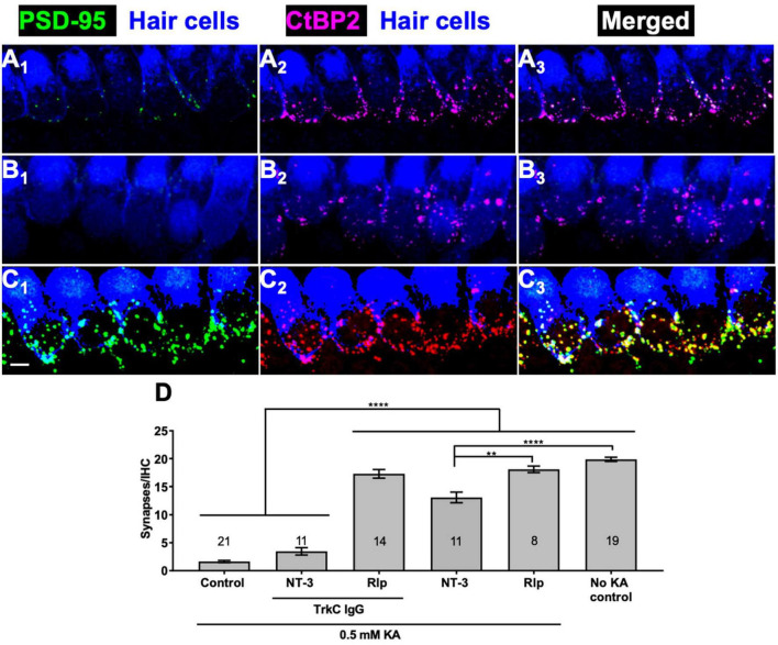
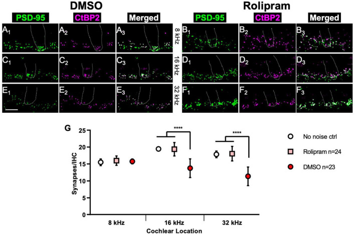
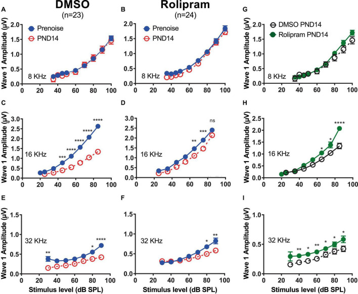
Results: We showed that the application of the cell membrane-permeable cAMP agonist 8-cpt-cAMP or the cAMP phosphodiesterase inhibitor rolipram promotes significant regeneration of synapses in vitro within twelve hours after their destruction by KA. This is independent of neurotrophin-3, which also promotes synapse regeneration. Moreover, of the two independent signaling effectors activated by cAMP - the cAMP Exchange Protein Activated by cAMP and the cAMP-dependent protein kinase - it is the latter that mediates synapse regeneration. Finally, we showed that systemic delivery of rolipram promotes synapse regeneration in vivo following NICS.

Discussion: In vitro experiments show that cAMP signaling promotes synapse regeneration after excitotoxic destruction of cochlear synapses and does so via PKA signaling. The cAMP phosphodiesterase inhibitor rolipram promotes synapse regeneration in vivo in noise-exposed mice. Systemic administration of rolipram or similar compounds appears to provide a minimally invasive therapeutic approach to reversing synaptopathy post-noise.

Keywords: auditory nerve; cochlea; cyclic AMP-dependent protein kinase; noise; regeneration; spiral ganglion neuron; synapse; synaptopathy.


Conflict of interest statement

The authors declare that the research was conducted in the absence of any commercial or financial relationships that could be construed as a potential conflict of interest. The author(s) declared that they were an editorial board member of Frontiers, at the time of submission. This had no impact on the peer review process and the final decision.

Figures

FIGURE 1
FIGURE 1
Cyclic AMP-dependent protein kinase signaling promotes synapse regeneration. (A–F) Examples of organotypic cochlear cultures: middle region of P5 rat cochleae exposed to 0.5 mM KA for 2 h and fixed after an additional 72 h post-KA exposed to the indicated experimental treatments: (A) no kainate exposure control; (B–F) 0.5 mM kainate; (B) control, no added factors; (C) 1 mM cpt-cAMP; (D) 1 mM cpt-cAMP with 20 μM H-89; (E) 0.5 mM 100 μM 8-cpt 2MeO cAMP; (F) 2.4 μM rolipram. In this figure and all following figures showing confocal images, what is shown in each image is a projection of the 3D volume to a single plane (Z-projection), the actual synapse counts were done on the original 3D confocal image stacks. The explants were labeled to detect postsynaptic densities (PSDs, anti-PSD95), presynaptic ribbons (anti-CtBP2), and hair cells (anti-myosin VI). Column 1 shows PSDs (green) and hair cell labeling (blue); column 2 shows ribbons (magenta) and hair cell labeling (blue); column 3 shows merged hair cell, ribbon and PSD labeling to show colocalization of PSDs and ribbons. Scale is the same for all images; scale bar, 20 μm (panel F1). (G) Synapses/IHC present 72 h after a 2 h exposure to KA, relative to cultures not exposed to KA (expressed as a percentage). Shown are means ± SEM for the indicated number of organotypic cochlear explant cultures. Significance of differences among conditions for each value of kainate was determined by ANOVA with Tukey’s multiple comparisons test: *p < 0.05, **p < 0.01, ****p < 0.0001. After KA, cultures were maintained in medium containing CPT-cAMP (1 mM), CPT-cAMP (1 mM) and PKA inhibitor H89 (20 μM), EPAC-selective activator 8-pCPT-2-O-Me-cAMP (100 μM), or PDE4 inhibitor rolipram (Rlp) at the three concentrations indicated.
FIGURE 2
FIGURE 2
Rolipram promotes synapse regeneration within 12 h. Explants were labeled to detect SGN peripheral axons, postsynaptic densities, presynaptic ribbons, and hair cells and representative images displayed as described in Figure 1. (A) No KA control; (B–E) 2 h exposure to 0.5 mM KA followed by 16 h in control medium with no added factors (B) or in medium with 2.4 μM rolipram for 8 h (C) or 12 h (D) or 16 h (E). All panels are at the same magnification. Scale is the same for all images; scale bar, 20 μm (panel E1). (F) Quantitation of synapses/IHC present 8, 12, or 16 h after a 2 h exposure to KA for cultures maintained with or without rolipram (2.4 μM). Shown are means ± SEM for the indicated number of organotypic cochlear explant cultures. The significance of differences among conditions was determined by ANOVA with Tukey’s multiple comparisons tests: ****p < 0.0001.
FIGURE 3
FIGURE 3
Rolipram-induced synapse regeneration does not require endogenous NT-3. TrkC-IgG was used to block TrkC signaling. Explants were labeled to detect postsynaptic densities, presynaptic ribbons, and hair cells and representative images displayed as described in Figure 1. Synapses were allowed to regenerate for 12 h after KA exposure in the indicated conditions. (A) NT-3 (50 ng/ml); (B) NT-3 (50 ng/ml), and TrkC-IgG (2 μg/ml); (C) rolipram (Rlp, 2.4 μM) and TrkC-IgG (2 μg/ml). All panels are at the same magnification. Scale is the same for all images; scale bar, 20 μm (lower left panel C1). (D) Quantification of synapses/IHC present 12 h after a 2 h exposure to KA for cultures maintained with NT-3 (50 ng/ml) or rolipram (2.4 μM) and with or without TrkC-IgG (2 μg/ml). Shown are means ± SEM for the indicated number of organotypic cochlear explant cultures. The significance of differences among conditions was determined by ANOVA with Tukey’s multiple comparisons test: **p < 0.01, ****p < 0.0001.
FIGURE 4
FIGURE 4
In vivo assessment of rolipram: timeline of ABR measures and corresponding auditory thresholds. (A) Timeline for the noise exposure experiments shows times relative to noise exposure (day 0) for three ABR measures: (1) prenoise, to verify normal hearing and establish a baseline for each individual mouse for normalization of post-noise measures; (2) postnoise day 1 (PND1), to measure temporary threshold shift (TTS) and verify effective noise exposure; and (3) postnoise day 14 (PND14) to ensure lack of permanent threshold shift and to measure wave-I amplitude. (B–D) Measures of ABR thresholds (mean ± SD) at, respectively, 8, 16, and 32 kHz for each of the three timepoints shown in panel (A) for noise-exposed mice implanted with subcutaneous minipumps delivering systemically only DMSO (DMSO, n = 23) or rolipram in DMSO (rolipram, n = 24). These data show a similar threshold elevation (>40 dB for 16 and 32 kHz tone pips) between prenoise and 1 day postnoise (TTS), and subsequent return to prenoise threshold by post-noise day 14. There was no significant difference in these measures between DMSO and rolipram, indicating that rolipram affects neither the TTS nor recovery from the TTS.
FIGURE 5
FIGURE 5
Mice treated with rolipram during noise exposure exhibit reduced synapse loss. (A–F) Dissected cochlear wholemount preparations were labeled (see section “Materials and methods”) to detect postsynaptic densities (green) and presynaptic ribbons (magenta), using the same antibodies as for Figure 1. Shown are representative examples at three cochlear locations, 8 kHz (A,B), 16 kHz (C,D), and 32 kHz (E,F) from mice in which either control DMSO (A,C,E) or DMSO with 4.4 mg/Kg/day rolipram (B,D,F) was delivered starting after the noise exposure. The location of a hair cell is shown by the dotted outline in each panel. Scale is the same for all images; scale bar, 10 μm (lower left panel E1). (G) Quantification of synapses/IHC for noise-exposed mice treated with control DMSO vehicle only and noise-exposed mice treated with rolipram in DMSO. Shown are means ± SEM for the indicated number of ears. The significance of differences among the three experimental groups was determined by ANOVA with Tukey’s multiple comparisons test: ****p < 0.0001. Synapses/IHC for control non-noise-exposed ears (No noise ctrl) is from Hu et al. (2020).
FIGURE 6
FIGURE 6
Mice treated with rolipram during noise exposure exhibit reduced decline in wave-I amplitude. (A–F) Measurements of ABR wave-I amplitude as a function of stimulus intensity (“growth curves”) made, on the same mice, prenoise (empty circle) and 14 days postnoise (PND14, filled circle) for 8 kHz (A,B), 16 kHz (C,D), and 32 kHz (E,F) tone pips, at indicated stimulus levels, for mice in which either control DMSO (A,C,E) or DMSO with rolipram (D–F) was delivered starting after the noise exposure. Shown are means ± SEM for the indicated number of ears. The curves were constructed by fitting the data (by least squares) to a second-order polynomial. Significance of amplitude differences between prenoise and PND14 measures at each stimulus level is as shown: *p < 0.05, **p < 0.01, ***p < 0.001, ****p < 0.0001. Repeated measure two-way ANOVA with Sidak’s multiple comparisons test for all suprathreshold stimulus levels and prenoise vs. PND14. (G–I) Direct comparison of PND14 wave-I amplitude between control DMSO and Rolipram groups. Significant differences between prenoise and PND14 were found at 16 and 32 kHz.
FIGURE 7
FIGURE 7
Mice chronically treated with rolipram for 2 weeks exhibit no significant changes in ABR wave-I amplitude or synapse number. (A) Direct comparison of wave-I amplitude measured, at 16 kHz, 1 day prior to rolipram (RLP) treatment (PreRLP) and after 2 weeks of rolipram treatment (PostRLP), assessed as summarized for Figure 6. Shown are means ± SEM. The curves were constructed by fitting the data (by least squares) to a second-order polynomial. An F-test was used to ask whether the pre-rolipram and 14 day rolipram data were better fit by a single curve (null hypothesis) or, alternatively, by two different curves. The conclusion (alpha = 0.05) was to accept the null hypothesis, indicating no significant difference caused by 2 weeks of rolipram exposure. (B) ABR wave-I amplitude was determined prior to, and after 2 weeks, of continuous rolipram treatment (RLP) or control vehicle only (DMSO). The amplitude after treatment was normalized to the amplitude prior to treatment. Shown is the mean normalized amplitude at each stimulus level for the indicated number of mice. (C) Quantification of synapses/IHC for male and female mice and mice treated with rolipram for 14 days (RLP). Shown are means ± SEM for the indicated number of ears at three cochlear locations, 8, 16, and 32 kHz. There was no significant difference between males and females and no significant effect of rolipram on synapse number (two-way ANOVA).

Similar articles

References

    1. Aglah C., Gordon T., Posse de Chaves E. I. (2008). cAMP promotes neurite outgrowth and extension through protein kinase A but independently of Erk activation in cultured rat motoneurons. Neuropharmacology 55 8–17. 10.1016/j.neuropharm.2008.04.005 - DOI - PubMed
    1. Bailey E. M., Green S. H. (2014). Postnatal expression of neurotrophic factors accessible to spiral ganglion neurons in the auditory system of adult hearing and deafened rats. J. Neurosci. 34 13110–13126. 10.1523/JNEUROSCI.1014-14.2014 - DOI - PMC - PubMed
    1. Bandtlow C. E. (2003). Regeneration in the central nervous system. Exp. Gerontol. 38 79–86. - PubMed
    1. Bok J., Zha X. M., Cho Y. S., Green S. H. (2003). An extranuclear locus of cAMP-dependent protein kinase action is necessary and sufficient for promotion of spiral ganglion neuronal survival by cAMP. J. Neurosci. 23 777–787. 10.1523/JNEUROSCI.23-03-00777.2003 - DOI - PMC - PubMed
    1. Bolger G., Michaeli T., Martins T., St John T., Steiner B., Rodgers L., et al. (1993). A family of human phosphodiesterases homologous to the dunce learning and memory gene product of Drosophila melanogaster are potential targets for antidepressant drugs. Mol. Cell Biol. 13 6558–6571. 10.1128/mcb.13.10.6558-6571.1993 - DOI - PMC - PubMed

LinkOut - more resources