Review
. 2024 May 1;14(20):14374-14391.
doi: 10.1039/d4ra01514b. eCollection 2024 Apr 25.

Recent progress of electrospun nanofibers as burning dressings

Affiliations
Review

Recent progress of electrospun nanofibers as burning dressings

Shengwei Zhang et al. RSC Adv. .

Abstract

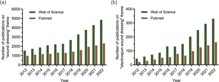
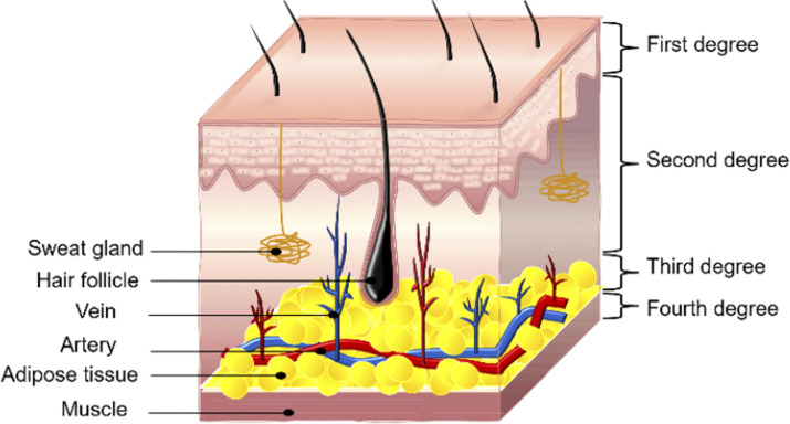
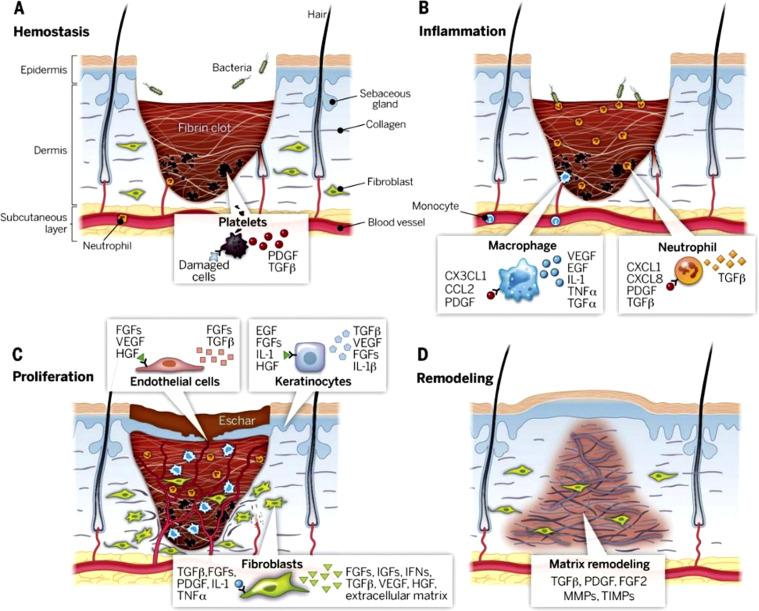
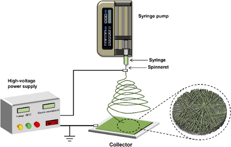
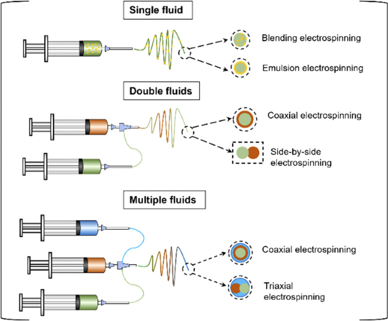
Burns are a global public health problem, which brings great challenges to public health and the economy. Severe burns often lead to systemic infection, shock, multiple organ failure, and even death. With the increasing demand for the therapeutic effect of burn wounds, traditional dressings have been unable to meet people's needs due to their single function and many side effects. In this context, electrospinning shows a great prospect on the way to open up advanced wound dressings that promote wound repairing and prevent infection. With its large specific surface area, high porosity, and similar to natural extracellular matrix (ECM), electrospun nanofibers can load drugs and accelerate wound healing. It provides a promising solution for the treatment and management of burn wounds. This review article introduces the concept of burn and the types of electrospun nanofibers, then summarizes the polymers used in electrospun nanofiber dressings. Finally, the drugs (plant extracts, small molecule drugs and nanoparticles) loaded with electrospun burn dressings are summarized. Some promising aspects for developing commercial electrospun burn dressings are proposed.


Conflict of interest statement

The authors declare no conflict of interest.

Figures

Fig. 1
Fig. 1. The literature related to the past ten years was searched in the databases of “PubMed” and “Web of Science”. (a) The number of literatures retrieved based on the theme “wound dressing”; (b) the number of literatures retrieved based on the theme “electrospun wound dressing”.
Fig. 2
Fig. 2. Burns depth and level. Burns are classified into four grades based on the size and depth of skin damage: first-degree, second-degree, third-degree, and fourth-degree burns.
Fig. 3
Fig. 3. Stages of wound healing: (A) hemostasis, (B) inflammation, (C) proliferation, (D) remodelling. Reproduced from ref. with permission from Science, copyright 2003.
Fig. 4
Fig. 4. Electrospinning device. The electrospinning device is mainly composed of an injection pump, a syringe connected with a spinneret, a high-voltage power supply, and a collector.
Fig. 5
Fig. 5. According to the number of fluids, electrospinning can be divided into single-fluid electrospinning (blended electrospinning and emulsion electrospinning), double-fluid electrospinning (coaxial electrospinning and side-by-side electrospinning), and multi-fluid electrospinning.
Fig. 6
Fig. 6. (a) Preparation process of the asymmetric wettable membrane. (b) The antibacterial and wound healing properties of asymmetric wettable film. Reproduced from ref. with permission from Wiley, copyright 2022.
Fig. 7
Fig. 7. (A) Silver sulfadiazine (SSD) loaded core–shell nanofibers: (a) SEM and TEM images of core–shell nanofibers (CSNF), 2% SSD loaded and 10% SSD loaded core–shell nanofibers (SSD-CSNF); (b) degradation profile of 2% SSD-CSNF; (c) the release profile of 2% and 10% SSD-CSNF. Adapted from ref. with permission from Elsevier, copyright 2022. (B) Poly(ethylene oxide)-chitosan nanofibers: (a) TEM image of ciprofloxacin-loaded nanofibers with a size of 100 nm; (b) cytotoxicity of nanofibers to human dermal fibroblasts and keratinocytes; (c) commutative drug release % vs. pH and time. Adapted from ref. with permission from Elsevier, copyright 2019. (C) Ginsenoside Rg3-loaded polyurethane/marine polysaccharide-based nanofiber dressings: (a) water absorption; (b) release curve of ginsenoside Rg3 with time; (c) and (d) are the effects of different concentrations of nanofiber dressing extracts on the proliferation of keratinocytes and fibroblasts. Adapted from ref. with permission from Elsevier, copyright 2023.
Fig. 8
Fig. 8. (A) Multifunctional nanofiber membranes containing black phosphorus/Rg1: (a) Escherichia coli and Staphylococcus aureus colonies remaining in the wound area on the third day; (b) image of umbilical vein endothelial cells in vitro wound healing experiment; (c) pictures of wounds treated with different materials on days 0, 3, 7 and 14; (d) H&E staining of wound tissues. Adapted from ref. with permission from Wiley, copyright 2022. (B) Core–shell structured anti-microbial nanofiber dressings (a) disk diffusion diagram of antibacterial activity of different core–shell nanofibers; (b) histological images of wounds untreated and treated with nanofiber dressings; (c) comparative images of wound treatment results at different time intervals. Adapted from ref. with permission from ACS, copyright 2021. (C) Application of core–shell nanofibers in burn treatment: (a) antibacterial effect of different core–shell nanofibers; (b) fluorescence microscope image of human dermal fibroblasts cultured on core–shell nanofibers; (c) histological sections of different groups on the 3rd and 7th day. Adapted from ref. with permission from Elsevier, copyright 2022.

Similar articles

Cited by

References

    1. Qi L. F. Zhang C. L. Wang B. Yin J. B. Yan S. F. Macromol. Biosci. 2022;22:e2100475. - PubMed
    1. Li Z. Y. Zhou F. Li Z. Y. Lin S. Y. Chen L. Liu L. X. Chen Y. M. ACS Appl. Mater. Interfaces. 2018;10:25194–25202. - PubMed
    1. Gholipour-Kanani A. Bahrami S. H. Rabbani S. IET Nanobiotechnol. 2016;10:1–7. - PMC - PubMed
    1. Chen M. M. Tian J. Liu Y. Cao H. Li R. Wang J. H. Wu J. L. Zhang Q. Q. Chem. Eng. J. 2019;373:413–424.
    1. Shpichka A. Butnaru D. Bezrukov E. A. Sukhanov R. B. Atala A. Burdukovskii V. Zhang Y. Y. Timashev P. Stem Cell Res. Ther. 2019;10:94. - PMC - PubMed