

Efficacy and Safety of High-Viscosity Bone Cement in Percutaneous Vertebroplasty for Kummell's Disease
- PMID: 38694703
- PMCID: PMC11058172
- DOI: 10.1007/s43465-024-01133-3
Efficacy and Safety of High-Viscosity Bone Cement in Percutaneous Vertebroplasty for Kummell's Disease
Abstract
Background: To analyze and evaluate the clinical outcomes of using high-viscosity bone cement compared to low-viscosity bone cement in percutaneous vertebroplasty (PVP) for treatment of Kummell's disease.
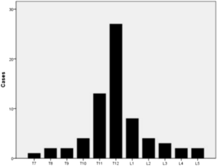
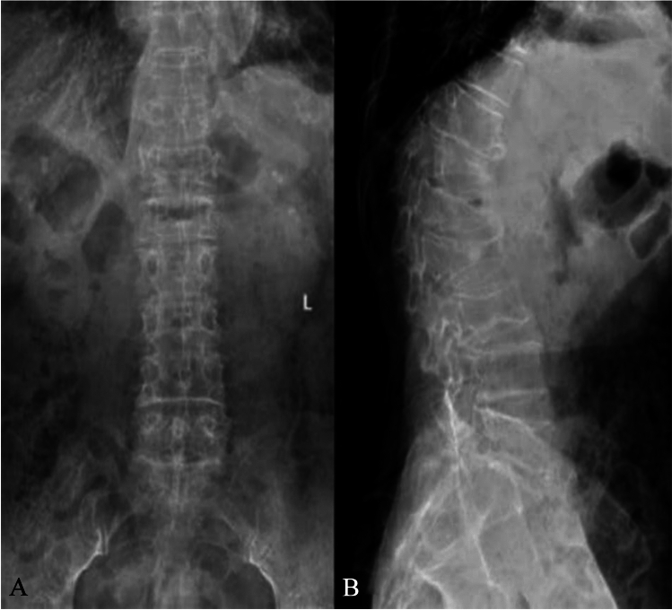
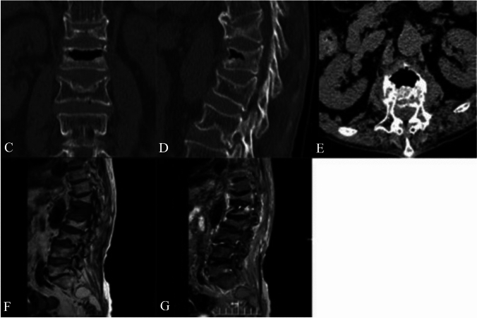
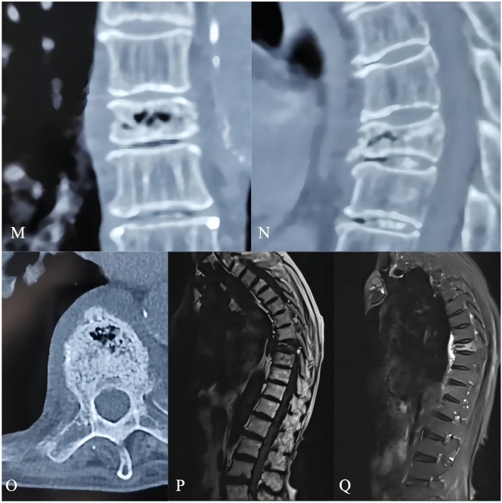
Methods: From July 2017 to July 2019, 68 Kummell's disease patients who underwent PVP were chosen and separated into 2 groups: H group (n = 34), were treated with high-viscosity bone cement and L group (n = 34), treated with low-viscosity bone cement during treatment. The operation time, number of fluoroscopy tests done, and amount of bone cement perfusion were recorded for both groups. Clinical outcomes were compared, by measuring their Visual Analog Scale (VAS), Oswestry Disability Index (ODI), Kyphosis Cobb's angle, vertebral height compression rate, and other complications.
Results: High-viscosity group showed less operation time and reduced number of fluoroscopy tests than the low-viscosity group (P < 0.05). When compared to preoperative period, both groups' VAS and ODI scores were significantly reduced at 1 day and 1 year postoperatively (P < 0.05). The vertebral height compression rate and Cobb's angle were significantly lower (P < 0.05) in both groups after surgery compared with those before surgery (P < 0.05). The cement leakage rate in group H was 26.5%, which was significantly lower than that in group L, which was 61.8% (P < 0.05).
Conclusions: High-viscosity and low-viscosity bone cement in PVP have similar clinical efficacy in reducing pain in patients during the treatment, but in contrast, high-viscosity bone cement shortens the operative time, reduces number of fluoroscopy views and vertebral cement leakage and improves surgical safety.
Keywords: Bone cement; Cement leakage; Compression fractures; High-viscosity; Kummell’s disease; Low-viscosity; Pulmonary embolism; Vertebroplasty.
© Indian Orthopaedics Association 2024. Springer Nature or its licensor (e.g. a society or other partner) holds exclusive rights to this article under a publishing agreement with the author(s) or other rightsholder(s); author self-archiving of the accepted manuscript version of this article is solely governed by the terms of such publishing agreement and applicable law.
Conflict of interest statement
Conflict of interestThe authors declare no conflict of interest.
Figures


References
-
- Kümmell H. Die rarefizierende ostitis der Wirbelkorper. Deutsche Med. 1895;21(1):180–181. doi: 10.1055/s-0029-1199707. - DOI
LinkOut - more resources
Full Text Sources