. 2024 Apr 16;7(8):9159-9166.
doi: 10.1021/acsanm.4c00668. eCollection 2024 Apr 26.

Biotinylated Photocleavable Semiconductor Colloidal Quantum Dot Supraparticle Microlaser

Affiliations

Biotinylated Photocleavable Semiconductor Colloidal Quantum Dot Supraparticle Microlaser

Charlotte J Eling et al. ACS Appl Nano Mater. .

Abstract

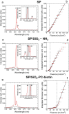
Luminescent supraparticles of colloidal semiconductor nanocrystals can act as microscopic lasers and are hugely attractive for biosensing, imaging, and drug delivery. However, biointerfacing these to increase functionality while retaining their main optical properties remains an unresolved challenge. Here, we propose and demonstrate red-emitting, silica-coated CdSxSe1-x/ZnS colloidal quantum dot supraparticles functionalized with a biotinylated photocleavable ligand. The success of each step of the synthesis is confirmed by scanning electron microscopy, energy dispersive X-ray and Fourier transform infrared spectroscopy, ζ-potential, and optical pumping measurements. The capture and release functionality of the supraparticle system is proven by binding to a neutravidin functionalized glass slide and subsequently cleaving off after UV-A irradiation. The biotinylated supraparticles still function as microlasers; e.g., a 9 μm diameter supraparticle has oscillating modes around 625 nm at a threshold of 58 mJ/cm2. This work is a first step toward using supraparticle lasers as enhanced labels for bionano applications.


Conflict of interest statement

The authors declare no competing financial interest.

Figures

Figure 1
Figure 1
(a) Schematic of oil-in-water emulsion technique used to synthesis SPs from CdSSe/ZnS CQDs. (b) Absorbance and PL emission spectra of CdSSe/ZnS CQDs. (c) Schematic of growth of silica shell using modified Stöber process and surface functionalization with biotinylated ligand. (d) SEM image of SP/SiO2-PC-biotin SPs.
Figure 2
Figure 2
(a) EDX spectra of SP and SP/SiO2-NH2. (b) FTIR spectra of supraparticle (SP), silica coated (SP/SiO2-NH2), and biotinylated SP (SP/SiO2-PC-biotin).
Figure 3
Figure 3
Schematic (not to scale) of the capture and release of biotinylated SPs. (a) The glass slide was functionalized with APTES and NHS-biotin with BSA added to aid nonspecific binding. Neutravidin was then washed over binding to the biotin. (b) The biotinylated SPs are then dropcast, leaving to bind for 3 min before washing. The sample is then irradiated under UV light. (c) The photocleavable groups in the ligand are activated under UV illumination, releasing the SPs which can then be washed away.
Figure 4
Figure 4
(a) Images of SP/SiO2-PC-biotin control (without neutravidin) and SP/SiO2-PC-biotin (with neutravidin) before and after UV irradiation. (b) Mean pixel intensity of images from (a). Description of the error bar calculations can be found in Suppporting Information.
Figure 5
Figure 5
(a, c, e) Emission spectra of a single SP at different pump fluences: (red) 81 mJ/cm2 and (black) 11 mJ/cm2 pump fluences, with a spectral resolution of 0.57 nm for (a) SP, (c) SP/SiO2-NH2, and (e) SP/SiO2-PC-biotin. Insets for (a), (c), (e): high spectral resolution (0.13 nm) emission spectra revealing substructure. Shaded areas denote areas used for analysis (M1, M4, and M7) for (b), (d), and (f). The diameters of the SPs were 8.9 ± 0.7 μm, 9.3 ± 0.3 μm, 9.6 ± 0.2 μm, respectively. (b), (d) and (f) are the corresponding laser transfer functions of SP, SP/SiO2-NH2, and SP/SiO2-PC-biotin. The thresholds are found to be 17.7 ± 1.7 mJ/cm2, 19.3 ± 1.8 mJ/cm2, and 58.1 ± 7.6 mJ/cm2, respectively. Further information on how the error bars were calculated can be found in Supporting Information.

References

    1. Yong K. T.; Law W. C.; Hu R.; Ye L.; Liu L.; Swihart M. T.; Prasad P. N. Nanotoxicity Assessment of Quantum Dots: From Cellular to Primate Studies. Chem. Soc. Rev. 2013, 42 (3), 1236–1250. 10.1039/C2CS35392J. - DOI - PubMed
    1. Foroozandeh P.; Aziz A. A. Insight into Cellular Uptake and Intracellular Trafficking of Nanoparticles. Nanoscale Res. Lett. 2018, 13, 339.10.1186/s11671-018-2728-6. - DOI - PMC - PubMed
    1. Brunetti V.; Chibli H.; Fiammengo R.; Galeone A.; Malvindi M. A.; Vecchio G.; Cingolani R.; Nadeau J. L.; Pompa P. P. InP/ZnS as a Safer Alternative to CdSe/ZnS Core/Shell Quantum Dots: In Vitro and in Vivo Toxicity Assessment. Nanoscale 2013, 5 (1), 307–317. 10.1039/C2NR33024E. - DOI - PubMed
    1. Zauner W.; Farrow N. A.; Haines A. M. R. In Vitro Uptake of Polystyrene Microspheres: Effect of Particle Size, Cell Line and Cell Density. J. Controlled Release 2001, 71, 39–51. 10.1016/S0168-3659(00)00358-8. - DOI - PubMed
    1. Zhao Z.; Ukidve A.; Kim J.; Mitragotri S. Targeting Strategies for Tissue-Specific Drug Delivery. Cell 2020, 181, 151–167. 10.1016/j.cell.2020.02.001. - DOI - PubMed

LinkOut - more resources