

Preliminary evaluation of an indwelling epidural catheter for repeat methylprednisolone administration in canine lumbosacral stenosis
- PMID: 38694734
- PMCID: PMC11017932
Preliminary evaluation of an indwelling epidural catheter for repeat methylprednisolone administration in canine lumbosacral stenosis
Abstract
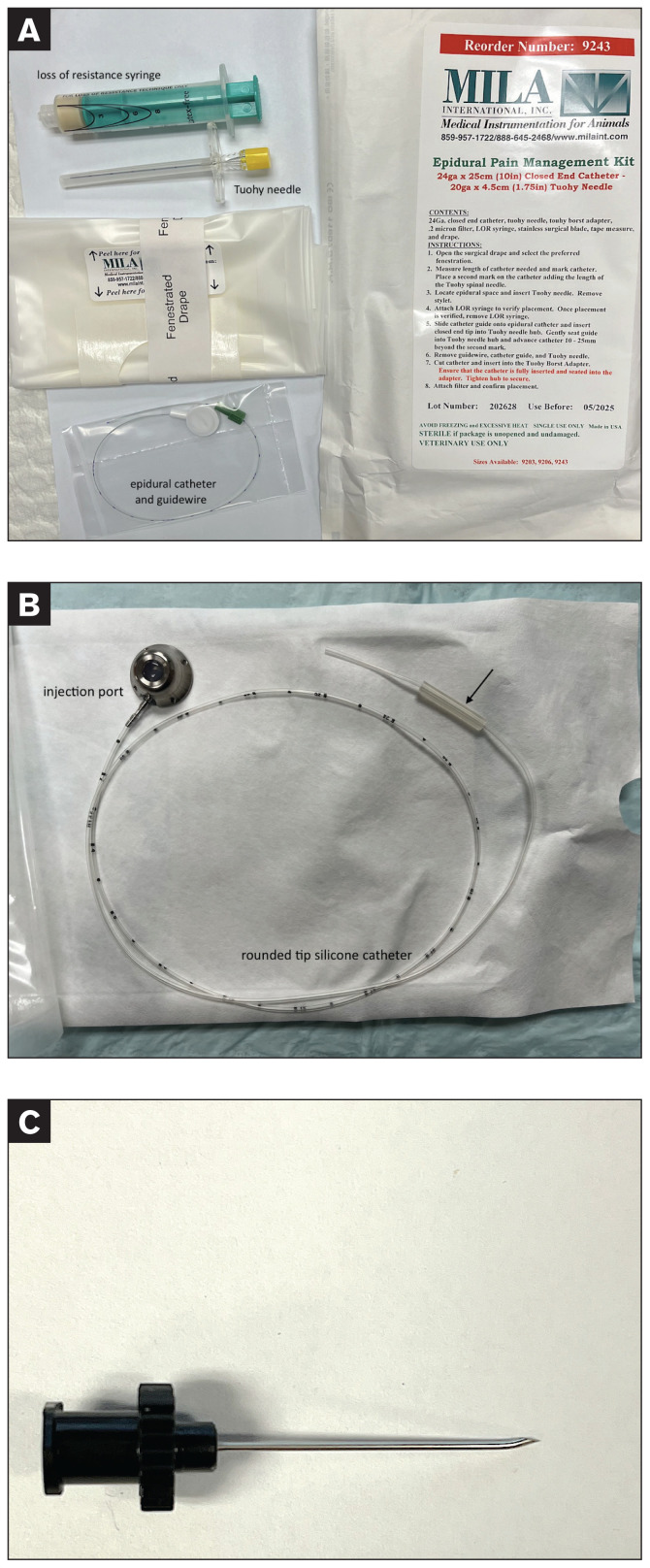
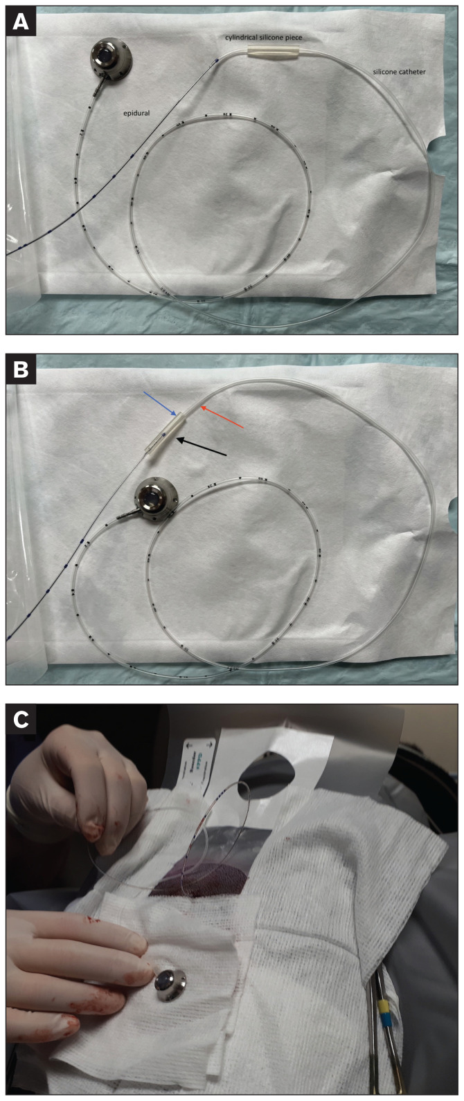
Objective: To determine the complications, outcomes, and patency of a permanent epidural catheter and subcutaneous access port system (ECAPS) as part of conservative management of degenerative lumbosacral stenosis in dogs.
Animals and procedure: Medical records of 11 client-owned dogs that underwent an ECAPS insertion were evaluated retrospectively. Clinical signs, complications related to the procedure, and system patency are reported.
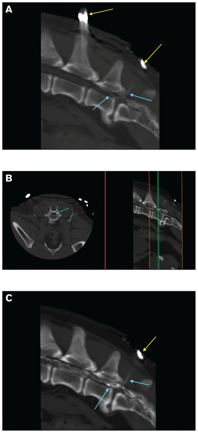
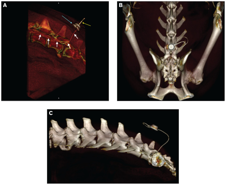
Results: All dogs had lumbosacral pain at their initial neurological assessment, with comfort levels adequately controlled following epidural infiltrations. None suffered from complications related to the ECAPS procedure. In 10 dogs, there were no malfunctions for the duration of the study. However, in 1 dog, there was a suspected leak at Day 814. The longest duration of patency reported in this study was 870 d (at the time of writing).
Conclusion: Placement of an ECAPS is a feasible technique and a viable option to permit repeated epidural injections of steroids in dogs with degenerative lumbosacral stenosis that is managed conservatively. Further studies are required to evaluate complication rates.
Évaluation préliminaire d’un cathéter épidural permanent (à demeure) pour l’administration répétée de méthylprednisolone lors de sténose lombosacrée dégénérative chez le chien.
Objectif: Décrire la technique, les complications, les résultats et la perméabilité d’un système composé d’un cathéter épidural et d’un port d’injection sous-cutanée (ECAPS) pour le traitement médical de la sténose lombosacrée dégénérative chez le chien.
Animaux et protocole: Les dossiers médicaux de 11 chiens appartenant à des clients ayant subi l’implantation d’un ECAPS ont été évalués de façon rétrospective. Cette étude décrit les signes cliniques, les complications reliées à la procédure et la perméabilité du système.
Résultats: Tous les patients inclus présentaient de la douleur lombosacrée à l’examen initial. Le niveau de confort de tous les patients suite aux injections épidurales fut maitrisé de façon adéquate. Aucun des patients n’a subi de complications reliées à l’implantation du système. Le système n’a pas démontré de dysfonctionnement dans le cas de dix patients. Chez un des patients, une fuite fut suspectée au jour 814. La durée maximale de perméabilité enregistrée dans cette étude est de 870 jours (au moment de la rédaction).
Conclusion: L’implantation d’un système ECAPS représente une option faisable et viable pour l’administration additionnelle de stéroïdes pour une gestion conservatrice de sténose lombosacrée dégénérative chez les chiens atteints. Des recherches supplémentaires sont requises pour l’évaluation des taux de complications.(Traduit par les auteurs).
Copyright and/or publishing rights held by the Canadian Veterinary Medical Association.
Figures

Similar articles
-
Amantadine as a therapeutic option for neuropathic pain in dogs with degenerative lumbosacral stenosis.BMC Vet Res. 2025 Jul 16;21(1):469. doi: 10.1186/s12917-025-04911-9. BMC Vet Res. 2025. PMID: 40671053 Free PMC article.
-
Prescription of Controlled Substances: Benefits and Risks.2025 Jul 6. In: StatPearls [Internet]. Treasure Island (FL): StatPearls Publishing; 2025 Jan–. 2025 Jul 6. In: StatPearls [Internet]. Treasure Island (FL): StatPearls Publishing; 2025 Jan–. PMID: 30726003 Free Books & Documents.
-
Indwelling bladder catheterisation as part of intraoperative and postoperative care for caesarean section.Cochrane Database Syst Rev. 2014 Apr 11;2014(4):CD010322. doi: 10.1002/14651858.CD010322.pub2. Cochrane Database Syst Rev. 2014. PMID: 24729285 Free PMC article.
-
Use of platelet transfusions prior to lumbar punctures or epidural anaesthesia for the prevention of complications in people with thrombocytopenia.Cochrane Database Syst Rev. 2018 Apr 30;4(4):CD011980. doi: 10.1002/14651858.CD011980.pub3. Cochrane Database Syst Rev. 2018. PMID: 29709077 Free PMC article.
-
Policies for replacing long-term indwelling urinary catheters in adults.Cochrane Database Syst Rev. 2016 Jul 26;7(7):CD011115. doi: 10.1002/14651858.CD011115.pub2. Cochrane Database Syst Rev. 2016. PMID: 27457774 Free PMC article.
References
-
- Janssens L, Beosier Y, Daems R. Lumbosacral degenerative stenosis in the dog: The results of epidural infiltration with methylprednisolone acetate: A retrospective study. Vet Comp Orthop Traumatol. 2009;22:486–491. - PubMed
-
- Danielski A, Bertran J, Fitzpatrick N. Management of degenerative lumbosacral disease in cats by dorsal laminectomy and lumbosacral stabilization. Vet Comp Orthop Traumatol. 2013;26:69–75. - PubMed
-
- Suwankong N, Meij BP, Voorhout G, deBoer AH, Hazewinkel HAW. Review and retrospective analysis of degenerative lumbosacral stenosis in 156 dogs treated by dorsal laminectomy. Vet Comp Orthop Traumatol. 2008;21:285–293. - PubMed
-
- De Risio L, Sharp NJ, Olby NJ, Muñana KR, Thomas WB. Predictors of outcome after dorsal decompressive laminectomy for degenerative lumbosacral stenosis in dogs: 69 cases (1987–1997) J Am Vet Med Assoc. 2001;219:624–628. - PubMed
MeSH terms
Substances
LinkOut - more resources
Full Text Sources
Medical