Review
. 2024 Apr 22:37:269-298.
doi: 10.1016/j.bioactmat.2024.03.030. eCollection 2024 Jul.

Biomaterial-enhanced treg cell immunotherapy: A promising approach for transplant medicine and autoimmune disease treatment

Affiliations
Review

Biomaterial-enhanced treg cell immunotherapy: A promising approach for transplant medicine and autoimmune disease treatment

Kazem Mashayekhi et al. Bioact Mater. .

Abstract

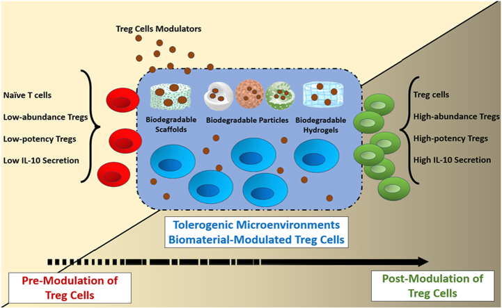
Regulatory T cells (Tregs) are crucial for preserving tolerance in the body, rendering Treg immunotherapy a promising treatment option for both organ transplants and autoimmune diseases. Presently, organ transplant recipients must undergo lifelong immunosuppression to prevent allograft rejection, while autoimmune disorders lack definitive cures. In the last years, there has been notable advancement in comprehending the biology of both antigen-specific and polyclonal Tregs. Clinical trials involving Tregs have demonstrated their safety and effectiveness. To maximize the efficacy of Treg immunotherapy, it is essential for these cells to migrate to specific target tissues, maintain stability within local organs, bolster their suppressive capabilities, and ensure their intended function's longevity. In pursuit of these goals, the utilization of biomaterials emerges as an attractive supportive strategy for Treg immunotherapy in addressing these challenges. As a result, the prospect of employing biomaterial-enhanced Treg immunotherapy holds tremendous promise as a treatment option for organ transplant recipients and individuals grappling with autoimmune diseases in the near future. This paper introduces strategies based on biomaterial-assisted Treg immunotherapy to enhance transplant medicine and autoimmune treatments.

Keywords: Autoimmune diseases; Biomaterials; Regulatory T cells; Transplantation.


Conflict of interest statement

No declaration for G.B.K.

Figures

Image 1
Graphical abstract
Fig. 1
Fig. 1
Treg development, differentiation, and the key factors: (A) Recognition of self-antigens in the thymus leads to the development of both tTregs and Tconv cells, which then migrate to lymphoid organs (B). (C) In peripheral organs, both tTregs and Tconv cells receive Treg-specific cytokines like IL-33, IL-2, and TGF-β, and encounter microbiota and their byproducts, leading to the formation of tTregs and pTregs pools in peripheral tissues. For example, pTregs originate from Tconv cells stimulated by RORγt + ILC3s and/or RORγt + DCs, along with Treg-specific cytokines in the gut. Additionally, CX3CR1+ DCs in the intestine can migrate to the thymus and present antigens from commensal microbiota to thymocytes, prompting their differentiation into tTregs. pTregs in peripheral tissues exhibit ST2+/RORγt+/GATA3-/Helios-//IL-10+, while tTregs in peripheral tissues are mostly ST2+/RORγt-/GATA3+/Helios+//IL-10+. (D) However, effector Tregs expressing ST2 migrate to peripheral organs, and their equilibrium is maintained via the IL-33/ST2 pathway. Different tissue-specific Tregs exhibit distinct key factors: gut Tregs demonstrate higher RORγt levels, adipose Tregs show elevated PPARγ, CNS Tregs express more Htr7, skin Tregs display higher RORα levels, and lung Tregs have increased IL4R and Notch4 expression. These characteristics are primarily observed in studies conducted on mice. Abbreviations arranged in alphabetical order: CX3CR1: CX3C motif chemokine receptor 1, DCs: Dendritic cells, GATA3: GATA Binding Protein 3, Helios: Transcription factor Ikzf2, HTR7: Serotonin receptor 7, IL-10: Interleukin-10, IL-2: Interleukin-2, IL-33: Interleukin-33, IL4R: IL-4 receptor, ILC3: Type-3 innate lymphoid cell, NOTCH4: Neurogenic locus notch homolog 4, PPARγ: Peroxisome proliferator-activated receptor gamma, pTregs: Peripheral-Derived Tregs, RORα: RAR-related orphan receptor alpha, RORγt: RAR-related orphan receptor gamma, ST2: IL-33 receptor, TGF-β: Transforming Growth Factor Beta, tTregs: Thymic-Derived Natural Tregs.
Fig. 2
Fig. 2
CNSs regions and related transcription factors: (A) T cell outer membrane: T cells express various transcription factors in response to different external stimulants like TGF-β, IL-2, vitamins D and C, retinoic acid, and TCR/CD28 stimulation, alongside internal regulators such as Tet enzymes. These factors bind to the FoxP3 promoter and enhancers, known as CNSs within the FoxP3 locus. (B) T cell nucleus: The FoxP3 locus comprises four CNS regions and 14 exons. These CNS regions enhance and regulate Foxp3 expression in response to downstream transcription factors (Shown with same colors) and the methylation status of the TSDR regions. The expressed Foxp3 protein acts as a master regulator transcription factor, promoting the expression of Treg-specific genes like GITR, CD25, CTLA-4, IL-2 suppression, and the maintenance of Foxp3 expression. (C) CNSs outcomes: Each CNS region responds to specific transcription factors and methylation statuses, thereby contributing to the generation of different Treg subsets such as tTregs, pTregs, and iTregs. Abbreviations arranged in alphabetical order: 5 hm C: 5-hydroxymethylcytosine, AP-1: Activator protein 1, ATF: Activating transcription factor, CBFβ: Core Binding Factor Beta, CNS: Conserved Noncoding Sequences, CREB: cAMP Response Element-Binding Protein, cRel: c-Rel homology Protein, CTLA4: Cytotoxic T-lymphocyte associated protein 4, DNMT3: DNA Methyltransferase 3, ETS1: Protein C-ets-1, Ex: Exon, Foxo: Forkhead box O, Foxp3: forkhead box P3, GITR: Glucocorticoid-induced TNFR-related protein, IL-2: Intelukin-2, IL-2R: Intelukin-2 receptor, iTregs: Induced Tregs, NFAT: Nuclear Factor of Activated T Cells, Nr4a: Nuclear Receptor Subfamily 4 A, PIAS1: Protein Inhibitor of Activated STAT1, Pol II: RNA polymerase II, pTregs: Peripheral-Derived Tregs, RAR: retinoic acid receptor, RunX1: Runt-related transcription factor 1, RXR: retinoid X receptor, Satb1: Special AT-rich sequence binding protein 1, Smad2/3/4: Smad transcription factors 2, 3, and 4, STAT5: Signal transducer and activator of transcription 5, TCR: T cell receptor, Tet: ten-eleven-translocation enzymes, TGF-β: Transforming Growth Factor Beta, TGF-βR: Transforming Growth Factor Beta Receptor, Tregs: Regulatory T cells, TSDR: Treg-specific demethylated region, tTregs: Thymic-Derived Natural Tregs, VDR: Vitamin D Receptor.
Fig. 3
Fig. 3
Controlled Release of Treg-Specific Signals: (A) Biomaterial-based platforms can control the release of specific-Treg signals (e.g., immunomodulators like Rapa, atRA, ITE, and Buty; cytokines such as IL-2 and TGF-β) over time, and they can also deliver auto- or allo-antigens alongside these signals (B), leading to the expansion of Tregs. (C) Biomaterial-based hydrogels and scaffolds, when functionalized with Treg modulators (e.g., FasL1, IL-2, IL-33, and TGF-β), can trigger tissue-specific Tregs in conjunction with self or allo cells. (D) In the dual-sized MP system, phagocytosable MPs are employed to deliver modulators (e.g., vitamin D3) and auto- or allo-antigens to DCs, while non-phagocytosable MPs are used for the controlled release of Treg modulators (e.g., TGF-β) and DC differentiation cytokines (e.g., GM-CSF), resulting in the expansion of Tol-DCs (E) and Tregs. Abbreviations arranged in alphabetical order: atRA: All-trans Retinoic Acid, Buty: Butyrate, D3: Vitamin D3, FasL: Fas Ligand, GM-CSF: Granulocyte-Macrophage Colony-Stimulating Factor, IL-10: Interleukin-10, IL-2: Interleukin-2, IL-33: Interleukin-33, ITE: 2-(1′Hindole-3′-carbonyl)-thiazole-4-carboxylic acid methyl ester, iTregs: Induced Tregs, MHC: Major Histocompatibility Complex, MPs: Microparticles, PD-1: Programmed cell death protein 1, PD-L1: Programmed death-ligand 1, pTregs: Peripheral-Derived Tregs, Rapa: Rapamycin, TCR: T Cell Receptor, TGF-β: Transforming Growth Factor Beta, Tol-DC: Tolerogenic Dendritic Cells, Tr1: Inducible T Regulatory Type 1, Treg: Regulatory T cells, tTregs: Thymic-Derived Natural Tregs.
Fig. 4
Fig. 4
Direct Functions of Biomaterials on Tregs Activation and Expansion: (A) The “backpack strategy" results in the activation of functionalized Tregs and, through a bystander mechanism, induces the conversion of circulatory T cells into Tregs. (B) Functionalized biomaterials equipped with anti-CD2/CD3/CD4/CD8 molecules deliver specific Treg signals (e.g., IL-2 and TGF-β) to T cells, thereby activating specific Treg subsets. (C) Biomaterial-based gene manipulation is employed to increase Treg gene expression of growth factors (e.g., IL-2 mRNA) or essential genes via miRNAs. This approach can be combined with Tregs' cytokines (e.g., IL-2 and TGF-β) to enhance the efficiency of Treg expansion. Abbreviations arranged in alphabetical order: IL-2: Interleukin-2, IL-2R: Interleukin-2 Receptor, MPs: Microparticles, TGF-β: Transforming Growth Factor Beta, Treg: Regulatory T cells.
Fig. 5
Fig. 5
Biomaterial-Induced Tol-Mic for Tregs: (A) Certain biomaterials possess inherent tolerogenic properties due to their unique physicochemical characteristics. When taken up by DCs, this leads to the differentiation of Tol-DCs. (B) Biomaterial-based delivery systems are employed to transport essential immunomodulators (e.g., vitamin D3 and IL-10) to Tol-DCs. These systems can also involve gene manipulation (e.g., encoding IL-10) to expand the population of Tol-DCs. (C) Biomaterial-based scaffolds are utilized to recruit T cells through chemoattractant cytokines like MCP-1 to the scaffold matrix. Subsequently, the differentiation of Tregs occurs through the interaction of modulatory molecules, such as Fas-FasL1, between Tregs and the scaffold. Simultaneously, the delivery of released auto- or allo-antigens to DCs results in the formation of a Tol-Mic and the expansion of antigen-specific Tregs. (D) The expanded Tol-DCs secrete anti-inflammatory cytokines like IL-10 and TGF-β, and they express immunomodulatory molecules such as FasL, PD-L1, and ICOS-L. These molecules, in turn, promote the expansion of Tregs. Abbreviations arranged in alphabetical order: CCR-1: CC chemokine receptor 1, FasL: Fas Ligand, ICOS: Inducible T-cell CO-Stimulator, ICOS-L: Inducible T-cell CO-Stimulator Ligand, IL-10: Interleukin-10, iTregs: Induced Tregs, MCP-1: Monocyte Chemoattractant Protein-1, MHC: Major Histocompatibility Complex, MPs: Microparticles, PD-1: Programmed cell death protein 1, PD-L1: Programmed death-ligand 1, pTregs: Peripheral-Derived Tregs, TCR: T Cell Receptor, TGF-β1: Transforming Growth Factor Beta 1, Tol-DC: Tolerogenic Dendritic Cells, Tol-Mic: Biomaterial-induced tolerogenic microenvironment, Tr1: Inducible T Regulatory Type 1, Tregs: Regulatory T cells, tTregs: Thymic-Derived Natural Tregs.
Fig. 6
Fig. 6
Biomaterials as Tol-aAPCs: (A) Biomaterials initiate signal-1 through p-MHC with or without signal-3, utilizing regulatory molecules like PD-L1, anti-Fas/FasL1, and CD47, or soluble modulators such as TGF-β. In the absence of signal-2 from co-stimulators like CD28, CD4 T cells either differentiate into Tregs or become anergic cells. (B) When biomaterials transduce signals-1 and 2 along with soluble signal-3 (e.g., TGF-β), this results in the differentiation of Tregs or induction of T cell anergy. (C) By crosslinking self/allo antigens with EDCI or SMCC onto syngeneic splenocytes or erythrocytes, antigens are presented to T cells with a disruption of MHC signals, ultimately leading to Treg differentiation (D). Abbreviations arranged in alphabetical order: aAPCs: Artificial APCs, ECDI: 1-ethyl-3-(3′-dimethylaminopropyl)-carbodiimide, ECM-Tol-aAPC: Engineered Cell Membrane-Derived Tol-aAPC, iTregs: Induced Tregs, p-MHC: Peptide-Major Histocompatibility Complex, PD-L1: Programmed death-ligand 1, pTregs: Peripheral-Derived Tregs, SMCC: Sulfosuccinimidyl-4-(N-maleimidomethyl) Cyclohexane-1-carboxylate, TCR: T Cell Receptor, TGF-β: Transforming Growth Factor Beta, Tol-aAPCs: Tolerogenic artificial Antigen-Presenting Cells, Tr1: Inducible T Regulatory Type 1, Tregs: Regulatory T cells.
Fig. 7
Fig. 7
Biomaterials to Facilitate Functional Tregs: (A) Gradual release of Tregs over time from biomaterial-based scaffolds, which are recruited to inflamed sites like transplanted organs, contributes to the alleviation of inflammation. Incorporating chemoattractant cytokines such as CCL-1 and IL-2 into Treg scaffolds enhances the effectiveness of implanted scaffolds (B). (C) Isolated Tregs with a low frequency can be expanded in vitro using MPs beads, leading to an increase in the efficiency of adoptive transfer Treg therapy (B). (D) By labeling expanded Tregs, it becomes possible to track the transferred cells over time and determine the fate of Tregs after adoptive transfer (B). Abbreviations arranged in alphabetical order: CCL-1: Chemokine ligand 1, CCR-8: CC chemokine receptor 8, IL-2: Interleukin-2, IL-2R: Interleukin-2 Receptor, MPs: Microparticles, Tregs: Regulatory T cells.
Fig. 8
Fig. 8
Immune Camouflage and Controlled Substrate Rigidity by Biomaterials: (A) PEG and its derivatives can effectively minimize or prevent host immune responses through PEG-based charge and steric camouflage, resulting in the inhibition of DCs, reduced levels of inflammatory cytokines (e.g., IL-1β, IL-2, TNF-α, and IFN-γ), expansion of Tregs, and an increase in anti-inflammatory cytokines (e.g., IL-10 and TGF-β). (B) Tissues/cells biotinylated for functionalization with streptavidin-modulators (e.g., PD-L1 and FasL1) can induce Tregs. (C) Biomaterials with varying stiffness are employed to optimize the expansion of Tregs, thereby increasing the efficiency of adoptive transfer Treg therapy. Abbreviations arranged in alphabetical order: DCs: Dendritic Cells, FasL: Fas Ligand, IFN-γ: Interferon Gamma, IL-10: Interleukin-10, IL-1β: Interleukin-1 beta, IL-2: Interleukin-2, PD-1: Programmed cell death protein 1, PD-L1: Programmed death-ligand 1, PEG: Polyethylene Glycol, TGF-β: Transforming Growth Factor Beta, TNF-α: Tumor Necrosis Factor Alpha, Tregs: Regulatory T cells.

Similar articles

Cited by

References

    1. Sakaguchi S., et al. Immunologic self-tolerance maintained by activated T cells expressing IL-2 receptor alpha-chains (CD25). Breakdown of a single mechanism of self-tolerance causes various autoimmune diseases. J. Immunol. 1995;155(3):1151–1164. - PubMed
    1. Liu W., et al. CD127 expression inversely correlates with FoxP3 and suppressive function of human CD4+ T reg cells. J. Exp. Med. 2006;203(7):1701–1711. - PMC - PubMed
    1. Friedman D.J., et al. The molecular Underpinnings of Treg plasticity in cancer. J. Immunol. 2023;210(1_Supplement):86.02.
    1. Bamidele A., et al. IL21 receptor-deficient regulatory T cells promote Resolution of intestinal inflammation by resisting metabolic Disturbance. J. Immunol. 2023;210(1_Supplement):66.06.
    1. Famili F., Wiekmeijer A.S., Staal F.J. The development of T cells from stem cells in mice and humans. Future Sci OA. 2017;3(3):Fso186. - PMC - PubMed