

Measuring uterine cavity volume with sonohysterography: A new objective method
- PMID: 38694830
- PMCID: PMC11060122
- DOI: 10.1177/1742271X231215502
Measuring uterine cavity volume with sonohysterography: A new objective method
Abstract
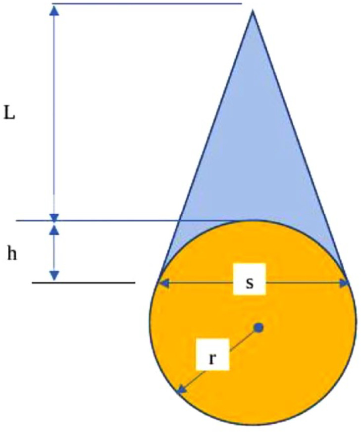
Introduction: The uterine cavity is a potential space with limited methods for evaluating its volume, limiting the evaluation of interventions' effectiveness in various uterine conditions. This study aims to objectively measure the uterine cavity volume using sonohysterography coupled with a Foley catheter to provide a normative model of age and parity-related uterine cavity volume.
Methods: The research included 35 women (group 1) with a total abdominal hysterectomy and 150 women (group 2) who underwent sonohysterography for various gynecologic indications. Saline infusion sonography was administered to all patients. The most common shape obtained after the saline infusion was taken to measure the uterine cavity's dimensions and volume. The uterine cavity volumes, as measured by sonohysterography, and the volumes of saline injected after the hysterectomy were compared.
Results: A significant association exists between uterine cavity volumes measured by sonohysterography and true volumes measured immediately after hysterectomy (p = 0.001). The association between uterine cavity volume measured by sonohysterography and using only a Foley catheter balloon was statistically insignificant (p = 0.13). A statistically significant positive association was observed between the uterine cavity volume and the patient's age and parity (p ⩽ 0.05).
Conclusion: Measuring the uterine cavity volume using a paediatric Foley catheter balloon coupled with sonohysterography offers an objective approach to measuring a normal (without gross pathologies) uterus volume. This technique would improve the diagnostic accuracy and the management of women with distinct uterine cavity morphologies.
Keywords: Catheters; hysterectomy; sonohysterography; uterine cavity volume.
© The Author(s) 2024.
Conflict of interest statement
The author(s) declared no potential conflicts of interest with respect to the research, authorship and/or publication of this article.
Figures


References
-
- Applebaum M. Imagine, imaging, imagination, http://www.drapplebaum.com/
-
- Piiroinen O. Studies in diagnostic ultrasound. Size of the non-pregnant uterus in women of child-bearing age and uterine growth and foetal development in the first half of normal pregnancy. Acta Obstet Gynecol Scand 1975; 54: 3–60. - PubMed
-
- Sørnes T, Bakke T. Uterine size, parity and umbilical cord length. Acta Obstet Gynecol Scand 1989; 68: 439–441. - PubMed
LinkOut - more resources
Full Text Sources