. 2024 Jul 23;150(4):302-316.
doi: 10.1161/CIRCULATIONAHA.123.065304. Epub 2024 May 2.

Deficiency of the Deubiquitinase UCHL1 Attenuates Pulmonary Arterial Hypertension

Affiliations

Deficiency of the Deubiquitinase UCHL1 Attenuates Pulmonary Arterial Hypertension

Haiyang Tang et al. Circulation. .

Abstract

Background: The ubiquitin-proteasome system regulates protein degradation and the development of pulmonary arterial hypertension (PAH), but knowledge about the role of deubiquitinating enzymes in this process is limited. UCHL1 (ubiquitin carboxyl-terminal hydrolase 1), a deubiquitinase, has been shown to reduce AKT1 (AKT serine/threonine kinase 1) degradation, resulting in higher levels. Given that AKT1 is pathological in pulmonary hypertension, we hypothesized that UCHL1 deficiency attenuates PAH development by means of reductions in AKT1.

Methods: Tissues from animal pulmonary hypertension models as well as human pulmonary artery endothelial cells from patients with PAH exhibited increased vascular UCHL1 staining and protein expression. Exposure to LDN57444, a UCHL1-specific inhibitor, reduced human pulmonary artery endothelial cell and smooth muscle cell proliferation. Across 3 preclinical PAH models, LDN57444-exposed animals, Uchl1 knockout rats (Uchl1-/-), and conditional Uchl1 knockout mice (Tie2Cre-Uchl1fl/fl) demonstrated reduced right ventricular hypertrophy, right ventricular systolic pressures, and obliterative vascular remodeling. Lungs and pulmonary artery endothelial cells isolated from Uchl1-/- animals exhibited reduced total and activated Akt with increased ubiquitinated Akt levels. UCHL1-silenced human pulmonary artery endothelial cells displayed reduced lysine(K)63-linked and increased K48-linked AKT1 levels.

Results: Supporting experimental data, we found that rs9321, a variant in a GC-enriched region of the UCHL1 gene, is associated with reduced methylation (n=5133), increased UCHL1 gene expression in lungs (n=815), and reduced cardiac index in patients (n=796). In addition, Gadd45α (an established demethylating gene) knockout mice (Gadd45α-/-) exhibited reduced lung vascular UCHL1 and AKT1 expression along with attenuated hypoxic pulmonary hypertension.

Conclusions: Our findings suggest that UCHL1 deficiency results in PAH attenuation by means of reduced AKT1, highlighting a novel therapeutic pathway in PAH.

Keywords: models, animal; pulmonary arterial hypertension; ubiquitination.


Conflict of interest statement

None.

Figures
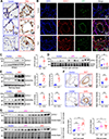
Figure 1.
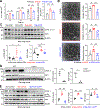
Figure 1.. Elevated UCHL1 expression in PAH.
A) Immunohistochemistry (IHC) reveals increased vascular-specific staining of UCHL1 in patients with PAH (n=3) compared to control patients (n=3). B) Immunofluorescence reveals co-localization of UCHL1 staining with vWF+ cells, consistent with increased expression in HPAECs (n=3). C) UCHL1 protein levels are higher in HPAECs isolated from subjects with PAH (n=5) than from controls (n=4). D) UCHL1 protein levels are higher in RV tissues isolated from subjects with RV hypertrophy (compensated RV or cRV with n=12 and decompensated RV or dRV with n=11) than from controls (n=17). E) Western blots demonstrate increased UCHL1 and GADD45α levels in MCT-exposed rats and in SuHx-exposed rats compared to their respective controls. F) IHC display increased UCHL1 staining in MCT-exposed rats and SuHx-exposed rats compared to their respective controls. G) UCHL1 protein levels are higher in RV tissues isolated from MCT-exposed rats (cRV and dRV, n=20 total) than from controls (n=10) and from PAB-exposed rats (cRV and dRV with n=17 total) than from controls (n=7). *p≤0.05, **p<0.01, ***p<0.001
Figure 2.
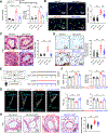
Figure 2.. UCHL1 inhibition reduces PAEC proliferation, tube formation, and Akt1 levels.
A) After VEGF administration (10 ng/mL), quantitative measurements of cell counts reveal reduced HPAEC proliferation over time with co-administration of LDN57444. BrdU incorporation assay reveals reduced cell proliferation in vitro with co-administration of LDN57444 in HPAECs exposed to VEGF. B) Using a tube formation assay, HPAECs exposed to LDN57444 exhibited significantly suppressed VEGF-induced tube formation due to formation of lower numbers of node, junction, and intervals observed in 3D culture. C) While total Akt1 levels rise mildly, hypoxia exposure significantly upregulates phospho-Akt protein levels in mouse lungs of WT mice. With LDN57444 administration, both total and phospho-Akt protein levels are reduced. D) Uchl1−/− rats demonstrate reduced p-Akt1 and slightly reduced total Akt1 levels in whole lung homogenates than WT. E) Tie2Cre-Uchl1fl/fl mice displayed reduced UCHL1, p-Akt1 and slightly reduced total Akt1 levels in whole lung homogenates compared to cage controls. The numbers of experiments (n) for each group are indicated in each bar. Analysis of variance, *p≤0.05, **p<0.01, ***p<0.001
Figure 3.
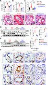
Figure 3.. Uchl1 deletion attenuates animal PH.
Rats with Uchl1 deletion resulted in reduced A) RVSP (Veh-WT: 27.0 α 1.0, Veh-Uchl1−/−: 23.9 α 0.6, MCT-WT: 83.6 α 5.6, MCT-Uchl1−/−: 40.4 α 6.1 mm Hg), and RVH (Veh-WT: 0.23 α 0.02, Veh-Uchl1−/−: 0.24 α 0.03, MCT-WT: 0.53 α 0.03, MCT-Uchl1−/−: 0.32 α 0.02), B and C) pulmonary artery medial histology and thickness, muscularized vessel counts and staining, as well as D) ki-67 staining in rat lungs compared to WT after exposure to MCT. E) Schematic strategy for the generation of Tie2Cre-Uchl1fl/fl mice and modeling in hypoxia. F) Summarized data showing RVSP and the Fulton index in control and Tie2Cre-Uchl1fl/fl mice exposed to normoxia and hypoxia. G) Representative images of pulmonary outflow pulse wave Doppler tracing and summarized data of echocardiography showing pulmonary acceleration time divided by pulmonary ejection time (PAT/PET ratios). H) Representative histology images of small PA and summarized data showing PA wall thickness in cage control and Tie2Cre-Uchl1fl/fl mice under normoxic and hypoxic conditions. Data above presented as mean ± SEM. *p≤0.05, **p<0.01, ***p<0.001. The numbers of experiments (n) for each group are indicated in each bar.
Figure 4.
Figure 4.. UCHL1-mediated linkage-specific modification of Akt1.
A) Immunoblot results revealed reduced total K48-linked protein levels in PAECs isolated from MCT-exposed rats compared to those from vehicle. B) Immunoblot results revealed increased K48-linked protein levels in whole lungs of Uchl1 knockout rats compared to those from cage controls or WT rats with MCT exposure. This trend was also observed in vehicle-exposed rats but was not statistically significant. C) Silencing UCHL1 for 48 hours in HPAECs resulted in increased K48-linked proteins which was further increased by exposure to MG132. Silencing UCHL1 for 48 hours in HPAECs resulted in increased K48-linked Akt1 (D) and reduced K63-linked Akt1 levels (E). Total loaded immunoprecipitated protein levels and actin levels in the unbound (to the immunoprecipitate) protein remained unchanged. * p≤0.05, **p<0.01, *** p<0.001.
Figure 5.
Figure 5.. UCHL1 epigenetics in patients with PAH.
A) The illustration depicts a highly GC-enriched region of the 5’ UCHL1 gene locus; rs9321 resides in this region. B) Analyses of GTeX reveals cis-eQTL of rs9321 with UCHL1 gene expression, with the T allele of associated with increased UCHL1 gene expression in lungs (p = 1.1 × 10⁻⁸). C) Association between rs9321 and cardiac index was statistically significant (P = 0.024). Each copy of T allele decreases cardiac index by a Beta of 0.2.
Figure 6.
Figure 6.. Gadd45α deletion results in attenuated murine hypoxic PH with reduced UCHL1 and Akt.
A) While both WT and Gadd45α−/− mice exhibit elevated RVSP, Gadd45α−/− mice demonstrate reduced RVSP (Nor-WT: 22.7 α 1.0, Hyp-WT: 41.8 α 0.8, Nor-Gadd45α−/−: 27.9 α 1.3, Hyp-Gadd45α−/−: 31.8 α 1.1 mmHg) and B) RVH (Nor-WT: 0.20 α 0.02, Hyp-WT: 0.49 α 0.03, Nor-Gadd45α−/−: 0.22 α 0.03, Hyp-Gadd45α−/−: 0.30 α 0.02) compared to WT mice when exposed to hypoxia. Data presented as mean ± SEM. The numbers of experiments (n) for each group are indicated in each bar. C) While both WT and Gadd45α−/− mice develop vascular remodeling after exposure to hypoxic PH, Gadd45α−/− mice demonstrate reduced pulmonary artery medial thickness index compared to WT mice hypoxic lungs. D) Hypoxia exposure upregulates total and phospho-Akt protein levels in whole mouse lung homogenates of WT mice. With Gadd45α deficiency, both total and phospho-Akt protein levels are reduced. E) Western blot from lung homogenates from wild type and Gadd45α−/− mice show reduced expression of UCHL1 protein. F) Immunohistochemistry reveals increased UCHL1 staining in WT mice exposed to chronic hypoxia compared to normoxia mice, which appears to be predominantly within the endothelium. *p≤0.05, **p<0.01, ***p<0.001.

References

    1. Pauwaa S, Machado RF and Desai AA. Survival in pulmonary arterial hypertension: A brief review of registry data. Pulm Circ. 2011;1:430–1. - PMC - PubMed
    1. Benza RL, Gomberg-Maitland M, Miller DP, Frost A, Frantz RP, Foreman AJ, Badesch DB and McGoon MD. The REVEAL Registry risk score calculator in patients newly diagnosed with pulmonary arterial hypertension. Chest. 2012;141:354–62. - PubMed
    1. McLaughlin VV, Archer SL, Badesch DB, Barst RJ, Farber HW, Lindner JR, Mathier MA, McGoon MD, Park MH, Rosenson RS, Rubin LJ, Tapson VF, Varga J, Harrington RA, Anderson JL, Bates ER, Bridges CR, Eisenberg MJ, Ferrari VA, Grines CL, Hlatky MA, Jacobs AK, Kaul S, Lichtenberg RC, Moliterno DJ, Mukherjee D, Pohost GM, Schofield RS, Shubrooks SJ, Stein JH, Tracy CM, Weitz HH and Wesley DJ. ACCF/AHA 2009 expert consensus document on pulmonary hypertension: a report of the American College of Cardiology Foundation Task Force on Expert Consensus Documents and the American Heart Association: developed in collaboration with the American College of Chest Physicians, American Thoracic Society, Inc., and the Pulmonary Hypertension Association. Circulation. 2009;119:2250–94. - PubMed
    1. Wilkins MR. Pulmonary hypertension: the science behind the disease spectrum. Eur Respir Rev. 2012;21:19–26. - PMC - PubMed
    1. Rhodes CJ, Batai K, Bleda M, Haimel M, Southgate L, Germain M, Pauciulo MW, Hadinnapola C, Aman J, Girerd B, Arora A, Knight J, Hanscombe KB, Karnes JH, Kaakinen M, Gall H, Ulrich A, Harbaum L, Cebola I, Ferrer J, Lutz K, Swietlik EM, Ahmad F, Amouyel P, Archer SL, Argula R, Austin ED, Badesch D, Bakshi S, Barnett C, Benza R, Bhatt N, Bogaard HJ, Burger CD, Chakinala M, Church C, Coghlan JG, Condliffe R, Corris PA, Danesino C, Debette S, Elliott CG, Elwing J, Eyries M, Fortin T, Franke A, Frantz RP, Frost A, Garcia JGN, Ghio S, Ghofrani HA, Gibbs JSR, Harley J, He H, Hill NS, Hirsch R, Houweling AC, Howard LS, Ivy D, Kiely DG, Klinger J, Kovacs G, Lahm T, Laudes M, Machado RD, Ross RVM, Marsolo K, Martin LJ, Moledina S, Montani D, Nathan SD, Newnham M, Olschewski A, Olschewski H, Oudiz RJ, Ouwehand WH, Peacock AJ, Pepke-Zaba J, Rehman Z, Robbins I, Roden DM, Rosenzweig EB, Saydain G, Scelsi L, Schilz R, Seeger W, Shaffer CM, Simms RW, Simon M, Sitbon O, Suntharalingam J, Tang H, Tchourbanov AY, Thenappan T, Torres F, Toshner MR, Treacy CM, Vonk Noordegraaf A, Waisfisz Q, Walsworth AK, Walter RE, Wharton J, White RJ, Wilt J, Wort SJ, Yung D, Lawrie A, Humbert M, Soubrier F, Tregouet DA, Prokopenko I, Kittles R, Graf S, Nichols WC, Trembath RC, Desai AA, Morrell NW, Wilkins MR, Consortium UNBRD, Consortium UPCS and Consortium UPB. Genetic determinants of risk in pulmonary arterial hypertension: international genome-wide association studies and meta-analysis. Lancet Respir Med. 2018. - PMC - PubMed

MeSH terms