Review
. 2024 Jul 1;36(8):1620-1642.
doi: 10.1162/jocn_a_02177.

Contributions of Basal Ganglia Circuits to Perception, Attention, and Consciousness

Affiliations
Review

Contributions of Basal Ganglia Circuits to Perception, Attention, and Consciousness

Michelle J Redinbaugh et al. J Cogn Neurosci. .

Abstract

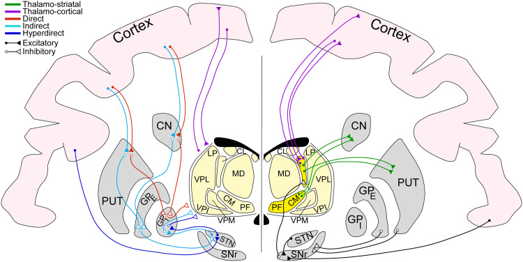
Research into ascending sensory pathways and cortical networks has generated detailed models of perception. These same cortical regions are strongly connected to subcortical structures, such as the basal ganglia (BG), which have been conceptualized as playing key roles in reinforcement learning and action selection. However, because the BG amasses experiential evidence from higher and lower levels of cortical hierarchies, as well as higher-order thalamus, it is well positioned to dynamically influence perception. Here, we review anatomical, functional, and clinical evidence to demonstrate how the BG can influence perceptual processing and conscious states. This depends on the integrative relationship between cortex, BG, and thalamus, which allows contributions to sensory gating, predictive processing, selective attention, and representation of the temporal structure of events.


Figures

<b>Figure 1.</b>
Figure 1.
Circuitry of the BG involves disinhibitory paths to the thalamus and direct thalamic paths to the striatum. (Left) Coronal slice demonstrating circuits between cortex (pink), BG (gray), and thalamus (light yellow) along direct (red), indirect (light blue), and hyperdirect (dark blue) pathways. Projections to and from thalamus reflect typical striato-thalamic, cortico-thalamic, or thalamo-cortical (purple) projections, applicable to multiple thalamic nuclei. Excitatory projections shown with closed ends; inhibitory shown with open white ends. (Right) Same conventions as left, but emphasizing connections of the intralaminar thalamic nuclei (dark yellow) and SNr: thalamo-striatal projections (green) and SNr connections (black). Note that all circuits shown are bilateral but are presented only in one hemisphere for visual clarity. Cortical projections indicate rough cortical depth (superficial, middle, or deep layers). Projections are general paths to the area and do not represent topography. GPe = external segment of the globus pallidus; PUT = putamen; LP = lateral posterior; VPM = ventroposterior medial; VPL = ventral posterolateral; VPI = ventroposterior inferior.
<b>Figure 2.</b>
Figure 2.
Parietal–striatal–thalamic circuits support consciousness states and DBS rescue of consciousness under general anesthesia. (A) Decoding accuracy of different conscious states (top; wake [W], sleep [S], and anesthesia [A]) and stimulation conditions (bottom; “Conscious Stim” refers to thalamic stimulations that increase consciousness under anesthesia and control stimulations that do not change consciousness) based on integrated information (Φ*) in a neural network consisting of frontal cortex, parietal cortex, CN, and thalamus. Note that thalamus was not included for stimulation conditions because electrode is used for stimulation. (B) Mean decrease in accuracy (MDA) for the models in A when system components, thalamus (T), CN (C), and superficial (s), middle (m), and deep (d) parietal (P) or frontal (F) cortical regions, are excluded during decoding. The greater the MDA, the greater the contribution of that region to decoding performance. Thalamus, CN, and deep parietal areas consistently contribute more to decoding accuracy. (C) Increase in neural complexity, Φ*, afforded by different system components for (top) conscious states and (bottom) during thalamic stimulations that induce consciousness in anesthetized macaques. CN contributions are just as high and often higher than most cortical regions, implying significant contributions to neural complexity. (D) Network diagrams showing the probability (grayscale of lines) of different brain areas (same as in B and C) associating in integrated structures when monkeys are (top left) awake, (top right) anesthetized, (bottom left) experiencing an increase in consciousness due to thalamic stimulation (Conscious Stim), or (bottom right) anesthetized, just before stimulations that induce consciousness. Monosynaptic anatomical pathways (Mono, in at least one direction) shown with thicker lines than multisynaptic projections (Multi). Red dashed lines show the minimum information partition (MIP), the way to “cut” the network that leads to minimum information loss. Conscious states, like Wake and Conscious stim, show a bias toward MIPs, which associate parietal and subcortical regions, indicating these structures contribute significantly to consciousness. Adapted with permission from Afrasiabi et al. (2021).
<b>Figure 3.</b>
Figure 3.
Altered parieto-striatal dynamics predominant in CL stimulation-induced perturbations of consciousness. (A) Decoding accuracy for Bayesian model trained on frontal, parietal, and striatal LFPs to distinguish between natural macaque stares and periods of vacant staring induced by CL DBS. Blue and red shading shows accuracy before and after event onset, respectively. Purple bar shows time window when decoding accuracy is significantly above chance. (B) Mean decrease in accuracy (MDA) for the decoding model in A attributed to each input feature. Features include superficial (s) or deep (d) layers of the FEF or lateral intraparietal area (LIP) and the CN at δ (1–4 Hz), θ (4–8 Hz), α (8–15 Hz), β (15–30 Hz), and γ (30–90 Hz) frequencies. MDA values for CN and LIP increase, leading to the onset of the vacant period, and remain strong during the period. (C) Normalized change in Φ* leading up to the onset of vacant periods induced by CL stimulation (red) or natural macaque staring (gray) for the same sessions decoded in A. The time course of changes in Φ* strongly resemble the time course of increased decoding accuracy. (D) For the same data in C, the strength of association between superficial (s) or deep (d) FEF (F) and striatal areas versus LIP (L) and striatal areas contributing to the integration measure, Φ*, approaching the onset of stares (gray bounded) or stimulation-induced (Stim-Induced) vacancy (red bounded). Decreases in Φ* coincide with a shift in integrated network structure, with CN dissociating from parietal cortex (both superficial and deep layers of LIP) and associating with frontal cortex (both superficial and deep layers of FEF). The time course of this change is similar to changes in the MDA attributed to parietal and striatal features of the decoding model in A. Adapted with permission from Redinbaugh et al. (2022). Sig = significance.

Similar articles

Cited by

References

    1. Aday, J. S., Wood, J. R., Bloesch, E. K., & Davoli, C. C. (2021). Psychedelic drugs and perception: A narrative review of the first era of research. Reviews in the Neurosciences, 32, 559–571. 10.1515/revneuro-2020-0094, - DOI - PubMed
    1. Adler, A., Katabi, S., Finkes, I., Israel, Z., Prut, Y., & Bergman, H. (2012). Temporal convergence of dynamic cell assemblies in the striato-pallidal network. Journal of Neuroscience, 32, 2473–2484. 10.1523/JNEUROSCI.4830-11.2012, - DOI - PMC - PubMed
    1. Afrasiabi, M., Redinbaugh, M. J., Phillips, J. M., Kambi, N. A., Mohanta, S., Raz, A., et al. (2021). Consciousness depends on integration between parietal cortex, striatum, and thalamus. Cell Systems, 12, 363–373. 10.1016/j.cels.2021.02.003, - DOI - PMC - PubMed
    1. Albantakis, L., Barbosa, L., Findlay, G., Grasso, M., Haun, A. M., Marshall, W., et al. (2023). Integrated information theory (IIT) 4.0: Formulating the properties of phenomenal existence in physical terms. PLoS Computational Biology, 19, e1011465. 10.1371/journal.pcbi.1011465, - DOI - PMC - PubMed
    1. Alexander, G. E., DeLong, M. R., & Strick, P. L. (1986). Parallel organization of functionally segregated circuits linking basal ganglia and cortex. Annual Review of Neuroscience, 9, 357–381. 10.1146/annurev.ne.09.030186.002041, - DOI - PubMed

Publication types