. 2024 May 2;29(1):9.
doi: 10.1007/s10911-024-09562-4.

A novel preclinical model of the normal human breast

Affiliations

A novel preclinical model of the normal human breast

Anthony J Wilby et al. J Mammary Gland Biol Neoplasia. .

Abstract

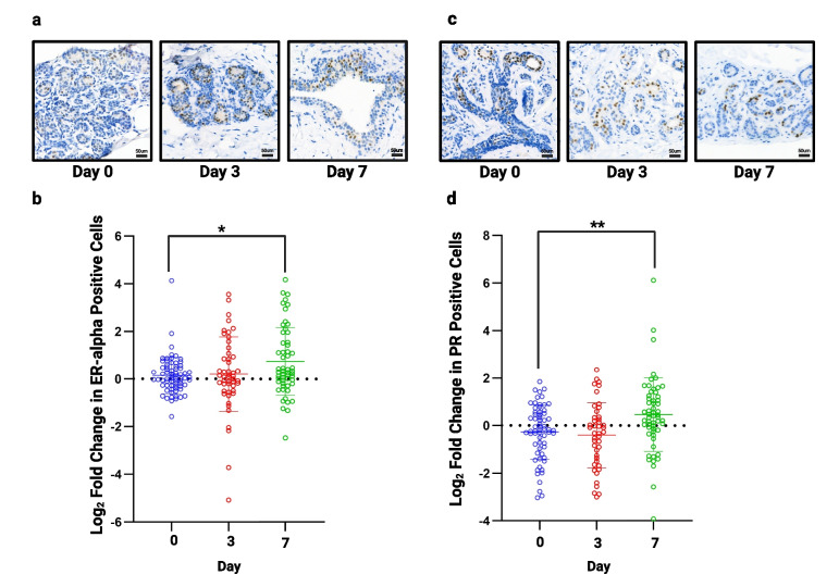
Improved screening and treatment have decreased breast cancer mortality, although incidence continues to rise. Women at increased risk of breast cancer can be offered risk reducing treatments, such as tamoxifen, but this has not been shown to reduce breast cancer mortality. New, more efficacious, risk-reducing agents are needed. The identification of novel candidates for prevention is hampered by a lack of good preclinical models. Current patient derived in vitro and in vivo models cannot fully recapitulate the complexities of the human tissue, lacking human extracellular matrix, stroma, and immune cells, all of which are known to influence therapy response. Here we describe a normal breast explant model utilising a tuneable hydrogel which maintains epithelial proliferation, hormone receptor expression, and residency of T cells and macrophages over 7 days. Unlike other organotypic tissue cultures which are often limited by hyper-proliferation, loss of hormone signalling, and short treatment windows (< 48h), our model shows that tissue remains viable over 7 days with none of these early changes. This offers a powerful and unique opportunity to model the normal breast and study changes in response to various risk factors, such as breast density and hormone exposure. Further validation of the model, using samples from patients undergoing preventive therapies, will hopefully confirm this to be a valuable tool, allowing us to test novel agents for breast cancer risk reduction preclinically.

Keywords: Explants; In vitro modelling; Normal breast; Prevention; Risk-reduction.


Conflict of interest statement

The authors declare no competing interests.

Figures

Fig. 1
Fig. 1
Explant model schematic: Individual steps are highlighted from tissue collection to fixing for immunohistochemistry. i. shows example of tissue processing, ii. tissue can be seen within the Boyden chamber, encased in hydrogel and iii. shows an example of the tissue microarray (TMA) produced for each sample
Fig. 2
Fig. 2
The effect of medium on proliferation. Following 3 and 7 days of culture in each of the media tested, tissue was fixed and assessed for proliferative rate using Ki67. a) shows representative images from each medium at each time point (TMA108). b) shows fold change in proliferation from day 0 in multiple cores from 4 patient samples. Significant increases in proliferation were seen in both Clevers’ medium (CM) and FCS medium at day 7. *P < 0.05 ** P < 0.01. Scale bar shows 50 µm
Fig. 3
Fig. 3
The effect of hydrogel support on proliferation. Following 3 and 7 days of culture in no hydrogel or each of the 3 hydrogel densities tested, tissue was fixed and assessed for proliferative rate using Ki67. a) shows representative images from each hydrogel support at each time point (TMA110). b) shows fold change in proliferation from day 0 in multiple cores from 3 patient samples. Significant increases in proliferation were seen when tissue was cultured with no support at day 7, within low support hydrogel at days 3 and 7 and in high support hydrogels at day 3. No change was seen using our moderate hydrogel (413.78 Pa). *P < 0.05 ** P < 0.01 *** P < 0.001. Scale bar shows 50 µm
Fig. 4
Fig. 4
Assessment of structure, proliferation and apoptosis in all samples. a) Representative images of H&E staining (TMA134) and b) Ki67 staining following 3 and 7 days of culture (TMA134) in optimised conditions. c) No significant change in proliferation was seen (n = 13). d) Representative images of caspase staining following 3 and 7 days of culture (TMA115), red arrow highlights single positive cell. Tissue cultured in FCS medium on day 3 used as an example of positive staining. Scale bar shows 50 µm
Fig. 5
Fig. 5
Assessment of hormone receptor expression in all samples. a) Representative images of ERα staining following 3 and 7 days of culture (TMA138). b) No significant change in ERα was seen at day 3 but there was a small, but significant increase at day 7 (n = 13). c) Representative images of PR staining following 3 and 7 days of culture (TMA134). d) No significant change in progesterone receptor was seen at day 3 but a small but significant, increase is seen at day 7 (n = 13). *P < 0.05 ** P < 0.01. Scale bar shows 50 µm
Fig. 6
Fig. 6
Assessment of oestrogen responsiveness. a) Proliferation was significantly increased at days 3 and 7 following the addition of 10 nM 17β-oestradiol. At day 3 proliferation remained significantly increased following the addition of 100 nM fulvestrant but at day 7, proliferation had fallen below control levels (day 7, no 17β-oestradiol). b) Progesterone receptor (PR) expression was significantly increased in the presence of 10 nM 17β-oestradiol at days 3 and 7 and this effect was blocked by 100 nM fulvestrant. *P < 0.05 **P < 0.01 ***P < 0.001
Fig. 7
Fig. 7
Assessment of immune cell infiltration. a) Representative images of CD4, CD8 and CD68 staining following 3 and 7 days of culture (TMA156). b) A significant decrease was seen in CD4 (n = 4) and CD8 (n = 7) cells after 7 days of culture and CD68 (n = 4) at 3 and 7 days. *P < 0.05 **P < 0.01. Scale bar shows 50 µm

Similar articles

Cited by

References

    1. Autier P, et al. Disparities in breast cancer mortality trends between 30 European countries: retrospective trend analysis of WHO mortality database. BMJ. 2010;341:c3620. doi: 10.1136/bmj.c3620. - DOI - PMC - PubMed
    1. Kohler BA, et al. Annual Report to the Nation on the Status of Cancer, 1975–2011, Featuring Incidence of Breast Cancer Subtypes by Race/Ethnicity, Poverty, and State. J Natl Cancer Inst. 2015;107(6):djv048. doi: 10.1093/jnci/djv048. - DOI - PMC - PubMed
    1. CRUK. Breast Cancer Statistics. 2021; Available from: https://www.cancerresearchuk.org/health-professional/cancer-statistics/s.... Accessed Mar 2024.
    1. Arnold M, et al. Current and future burden of breast cancer: Global statistics for 2020 and 2040. Breast. 2022;66:15–23. doi: 10.1016/j.breast.2022.08.010. - DOI - PMC - PubMed
    1. Weir HK, et al. The past, present, and future of cancer incidence in the United States: 1975 through 2020. Cancer. 2015;121(11):1827–1837. doi: 10.1002/cncr.29258. - DOI - PMC - PubMed

Publication types

LinkOut - more resources