. 2024 May 2;20(5):e1012125.
doi: 10.1371/journal.ppat.1012125. eCollection 2024 May.

Major Group-B Enterovirus populations deleted in the noncoding 5' region of genomic RNA modulate activation of the type I interferon pathway in cardiomyocytes and induce myocarditis

Affiliations

Major Group-B Enterovirus populations deleted in the noncoding 5' region of genomic RNA modulate activation of the type I interferon pathway in cardiomyocytes and induce myocarditis

Domitille Callon et al. PLoS Pathog. .

Abstract

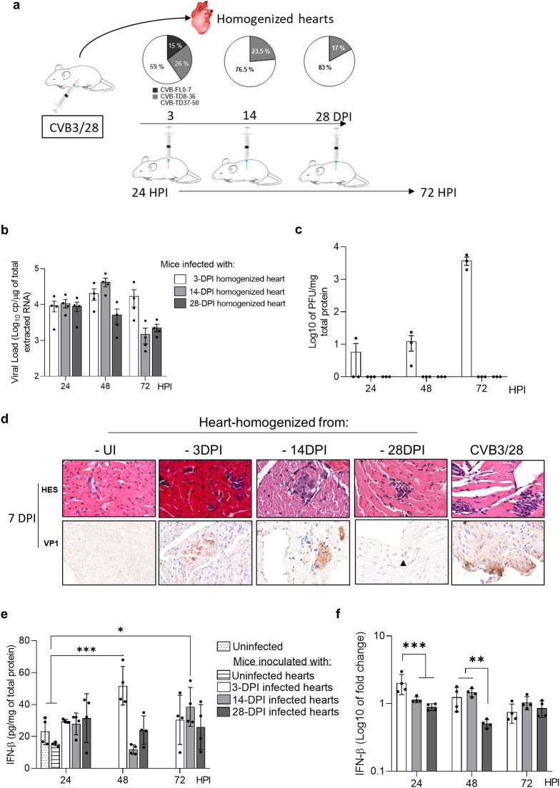
Major 5'-terminally deleted (5'TD) RNA forms of group-B coxsackievirus (CVB-5'TD) has been associated with myocarditis in both mice and humans. Although it is known that interferon-β (IFN-β) signaling is critical for an efficient innate immune response against CVB-induced myocarditis, the link between CVB-5'TD RNA forms and type I IFN signaling in cardiomyocytes remains to be explored. In a mouse model of CVB3/28-induced myocarditis, major early-emerging forms of CVB-5'TD RNA have been characterized as replicative viral populations that impair IFN-β production in the heart. Synthetic CVB3/28 RNA forms mimicking each of these major 5'TD virus populations were transfected in mice and have been shown to modulate innate immune responses in the heart and to induce myocarditis in mice. Remarkably, transfection of synthetic viral RNA with deletions in the secondary structures of the 5'-terminal CVB3 RNA domain I, modifying stem-loops "b", "c" or "d", were found to impair IFN-β production in human cardiomyocytes. In addition, the activation of innate immune response by Poly(I:C), was found to restore IFN-β production and to reduce the burden of CVB-5'TD RNA-forms in cardiac tissues, thereby reducing the mortality rate of infected mice. Overall, our results indicate that major early-emerging CVB3 populations deleted in the domain I of genomic RNA, in the 5' noncoding region, modulate the activation of the type I IFN pathway in cardiomyocytes and induce myocarditis in mice. These findings shed new light on the role of replicative CVB-5'TD RNA forms as key pathophysiological factors in CVB-induced human myocarditis.


Conflict of interest statement

The authors have declared that no competing interests exist.

Figures

Fig 1
Fig 1. The emergence of 5’terminally deleted (5’TD) CVB populations is associated with a low type I interferon response in the heart.
a Viral load levels (RT-qPCR) in the heart of infected mice at 1, 2, 3, 7, 14- and 28-days post-infection (DPI) (n = 4 to 9). b Viral titers (PFU) in the homogenized heart supernatants at 1, 2, 3, 7, 14- and 28 DPI (n = 4 to 9). c. Two-dimensional representation of the EV-B 5’non-coding region RNA sequences showing the position of the CVB3/28 5’ terminally deleted forms. d. CVB-5’TD and FL viral loads were assessed using a RACE-PCR method associated with a micro-electrophoresis, the profile of CVB-TD forms distribution was confirmed by NGS method, at 1, 2, 3, 7, 14- and 28-DPI (n = 4 to 9). Data represent mean ±SD. (Two-way ANOVA Multiple comparisons; **: p<0.01; ***: p<0.001). e Cytokines levels measured by ELISA (heatmap view) in homogenized heart supernatants expressed as fold change relative to uninfected mice data (n = 6). f IFN- β levels in the homogenized heart supernatants quantified by ELISA at indicated time points post-infection. g Linear regression curves and Spearman R coefficient of correlation between IFN-β levels and the proportions of 8-50nt 5’TD viral populations (2–3 DPI, n = 13). h Linear regression curves and Spearman R coefficient of correlation between IFN-β levels and total viral load (2–3 DPI, n = 13). i Full-length (white) and 5’terminally deleted (grey) viral forms proportions in hearts of CVB3/28 infected mice from 1 to 7 DPI. j Linear regression curves and Spearman R coefficient of correlation between cardiac IFN-β mRNA fold-changes and the 5’TD/FL ratio (24 to 72 HPI; n = 13). 5’TD: 5’terminally deleted; FL: full length; UI: uninfected.
Fig 2
Fig 2. Inoculation of cardiac CVB RNA populations in mice induces acute and chronic myocarditis.
a Schematic illustration of the experimentation. b Viral loads (RT-qPCR) in the hearts of mice infected with homogenized hearts or CVB3/28 (n = 4). c Viral titers (PFU assay) in the hearts of mice infected with homogenized hearts or CVB3/28. d Histology (HES) and immunohistochemistry for viral protein 1 (VP1, orange-brown staining) of mouse hearts at 7 DPI. e, f IFN-β cytokine and mRNA levels fold-changes (ELISA and RT-qPCR) in homogenized hearts supernatants of mice infected with homogenized hearts or CVB3/28 (n = 4) at the indicated points time post-infection. Data represent mean ± SD (Two-way ANOVA Multiple comparisons; *: p<0.05; **: p<0.01; ***: p<0.001 versus CVB3/28-infected mice or uninfected mice). UI: uninfected.
Fig 3
Fig 3. Major CVB-5’TD RNA forms transfection in DBA/2J mice induces acute myocarditis without IFN-β increase.
a Viral load (RT-qPCR) in the hearts of mice transfected with CVB-FL, 5’TD8, 5’TD21 or 5’TD50 RNA forms, at 8 hours post transfection (n = 4 to 6). b IFN-β cytokine levels (ELISA) in hearts of uninfected mice or mice transfected with vehicle alone, long-RNA, CVB-FL, 5’TD8, 5’TD21 or 5’TD50 RNA forms, at 8 hours post transfection. Data represent mean ± SEM (n = 4 to 8) (Two-way ANOVA Multiple comparisons; **: p<0.01; ns: non-significant; versus vehicle transfected mice). c Cytokines levels measured by ELISA (heatmap view) in homogenized heart supernatants expressed as fold change relative to uninfected mice data (n = 2 to 6). d Double-strands RNA quantification by ELISA assay in cardiac lysates between 24 and 72 hours post transfection. (n = 2 to 9) (Mann-Whitney U test: **: p<0.01). e Histological scoring of necrosis and inflammatory infiltrates in 3 slices per condition. f Histology (HES) and immunohistochemistry for viral protein 1 (VP1, orange-brown staining, black arrows) of mouse hearts at 7 DPI (n = 3). DPT: days post-transfection; UT: untransfected mice; TD: terminally deleted forms; VP1: Viral protein 1.
Fig 4
Fig 4. Impact of CVB3 viral populations on IFN-β pathway activation in cultured human cardiomyocytes.
a Schematic illustration of the experimentation: transfection of CVB-FL and CVB-5’TD (5’TD8, 5’TD15, 5’TD21 and 5’TD50) synthetic RNA forms into human cardiac myocytes cells (HCM). b Viral load (RT-qPCR) in HCM transfected cells with CVB-FL and CVB-5’TD (5’TD8, 5’TD15, 5’TD21 and 5’TD50) synthetic RNA at 8 hours post-transfection. c IFN-β cytokine levels (ELISA) in supernatants of HCM at 8 HPT of various CVB-TD/or full-length RNA forms. d Analysis of eIF4G cleavage, and levels of phosphorylated IRF3 (pS386-IRF3) and IRF3 on a Western blot prepared with HCM cells collected at 8 HPT. Poly(I:C) is used as positive control of IFN-β pathway activation. Data represent mean ± SD (n = 3) (Mann-Whitney U test; *: p<0.05; ****: p<0.0001 and ns: non-significant). eIF4G: eukaryotic translation initiation factor 4 G; HMW: high molecular weight Poly(I:C); IRF3: Interferon Regulatory Factor 3; eIF4G: eukaryotic initiation factor 4 gamma; UT: untransfected.
Fig 5
Fig 5. IFN-β signalling pathway stimulation by Poly(I:C) decreased 5’terminally deleted CVB3 RNA forms viral loads, cardiac lesions and mortality in DBA/2J mice.
a Survival curve and follow-up of body weight of CVB3/28-infected mice (n = 17; black square) compared to infected Poly(I:C) HMW-treated mice (n = 10; white circle) and uninfected mice (n = 9; black circle) (*: p<0.05 by Log-Rank test between infected and infected-treated groups). Data represent mean ±SEM (*: p<0.05, ***: p<0.001, by Tukey’s multiple comparison test between infected and infected-treated groups). b Semi-quantification of inflammation and necrosis in treated group (grey bar) and untreated group of mice (white bar) (n = 5 each) at 14 DPI. Data represent mean ± range (Mann-Whitney U test, *: p<0.05). c d IFN-β mRNA fold-changes and ELISA of IFN-β in homogenized heart supernatants of treated and untreated infected mice (n = 4 each) (Mann-Whitney U test, *: p<0.05). e f Viral titers (PFU assay) and loads (RT-qPCR) of treated infected mice versus untreated infected mice (n = 4) (Mann-Whitney U test, *: p<0.05). g CVB-TD and FL respective viral loads were assessed as previously described for treated infected mice (n = 3 each DPI) and compared with untreated infected mice (n = 6 to 10). Data represent mean ± SD. (Two-way ANOVA Multiple comparisons; *: p<0.05; untreated mice versus Poly(IC)-treated mice). HMW: high molecular weight poly(I:C). DPI: days post-infection.

Similar articles

Cited by

References

    1. Baggen J, Thibaut HJ, Strating JRPM, Kuppeveld FJM van. The life cycle of non-polio enteroviruses and how to target it. Nat Rev Microbiol. 2018; 16(6):368–381. doi: 10.1038/s41579-018-0005-4 - DOI - PubMed
    1. Genoni A, Canducci F, Rossi A, et al.. Revealing enterovirus infection in chronic human disorders: An integrated diagnostic approach. Sci Rep. 2017; 7(1):5013. doi: 10.1038/s41598-017-04993-y - DOI - PMC - PubMed
    1. Tracy S, Smithee S, Alhazmi A, Chapman N. Coxsackievirus can persist in murine pancreas by deletion of 5′ terminal genomic sequences: Coxsackievirus Persistence in the Pancreas. J Med Virol. 2015; 87(2):240–247. - PubMed
    1. Tam PE, Fontana DR, Messner RP. Coxsackievirus B1–induced chronic inflammatory myopathy: differences in induction of autoantibodies to muscle and nuclear antigens by cloned myopathic and amyopathic viruses. J Lab Clin Med. 2003; 142(3):196–204. doi: 10.1016/S0022-2143(03)00108-2 - DOI - PubMed
    1. Chapman NM, Kim K-S, Drescher KM, Oka K, Tracy S. 5’ terminal deletions in the genome of a coxsackievirus B2 strain occurred naturally in human heart. Virology. 2008; 375(2):480–491. doi: 10.1016/j.virol.2008.02.030 - DOI - PMC - PubMed

MeSH terms