. 2024 Jun:163:104252.
doi: 10.1016/j.jri.2024.104252. Epub 2024 Apr 29.

Individual disruption of 12 testis-enriched genes via the CRISPR/Cas9 system does not affect the fertility of male mice

Affiliations

Individual disruption of 12 testis-enriched genes via the CRISPR/Cas9 system does not affect the fertility of male mice

Akira Suzuki et al. J Reprod Immunol. 2024 Jun.

Abstract

More than 1200 genes have been shown in the database to be expressed predominantly in the mouse testes. Advances in genome editing technologies such as the CRISPR/Cas9 system have made it possible to create genetically engineered mice more rapidly and efficiently than with conventional methods, which can be utilized to screen genes essential for male fertility by knocking out testis-enriched genes. Finding such genes related to male fertility would not only help us understand the etiology of human infertility but also lead to the development of male contraceptives. In this study, we generated knockout mice for 12 genes (Acrv1, Adgrf3, Atp8b5, Cfap90, Cfap276, Fbxw5, Gm17266, Lrrd1, Mroh7, Nemp1, Spata45, and Trim36) that are expressed predominantly in the testis and examined the appearance and histological morphology of testes, sperm motility, and male fertility. Mating tests revealed that none of these genes is essential for male fertility at least individually. Notably, knockout mice for Gm17266 showed smaller testis size than the wild-type but did not exhibit reduced male fertility. Since 12 genes were not individually essential for male fertilization, it is unlikely that these genes could be the cause of infertility or contraceptive targets. It is better to focus on other essential genes because complementary genes to these 12 genes may exist.

Keywords: CRISPR/Cas9; Knockout mice; Male infertility; Spermatozoa; Testis.


Conflict of interest statement

Declaration of Competing Interest All authors declared no competing interests.

Figures
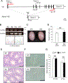
Fig. 1.
Fig. 1.
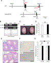
Phenotypic analysis of Nemp1 knockout male mice. (A) Genomic structure and knockout strategy of mouse Nemp1. Two gRNAs (red arrowheads) were designed to target the coding region (black boxes) in the first (exon 1) and the last exons (exon 9). Two primer sets (black arrows, Fw1 and Rv1; Fw2 and Rv2) were designed for gPCR. (B) Knockout and wild-type alleles were detected by gPCR using a pair of Fw1 and Rv1 and a pair of Fw2 and Rv2, respectively. Deletion of knockout allele was confirmed by Sanger sequencing. KO, He, and WT show homozygous knockout, heterozygous knockout, and wild-type mice, respectively. (C) Testis appearance of WT and Nemp1 KO mice. Scale bar, 2 mm. (D) Bar graphs show the average testicular weight of WT and Nemp1 KO mice. NS, no significant difference (P = 0.167). n, number of male mice in each. (E) Histological analysis of testes in WT and Nemp1 KO mice. Scale bar, 100 μm. (F) Morphology of spermatozoa collected from the cauda epididymides in WT and Nemp1 KO mice. Scale bar, 50 μm. (G) Bar graphs show percentages of motile spermatozoa in WT and Nemp1 KO mice. Sperm motility was analyzed at 10 min and 2 hrs after incubation in TYH medium. NS, no significant difference (P = 0.940 for 10 min, 0.815 for 2 hrs).
Fig. 2.
Fig. 2.
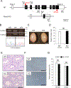
Phenotypic analysis of Spata45 knockout male mice. (A) Genomic structure and knockout strategy of mouse Spata45. Two gRNAs (red arrowheads) were designed in the introns before exon 2 and before exon 3 to disrupt exon 2. Coding regions are shown by black boxes. Three primers (black arrows, Fw1, Fw2, and Rv2) were designed for gPCR. (B) Knockout and wild-type alleles were detected by gPCR using a pair of Fw1 and Rv1 and a pair of Fw1 and Rv2, respectively. Deletion of knockout allele was confirmed by Sanger sequencing. KO, He, and WT show homozygous knockout, heterozygous knockout, and wild-type mice, respectively. (C) Testis appearance of WT and Spata45 KO mice. Scale bar, 2 mm. (D) Bar graphs show the average testicular weight of WT and Spata45 KO mice. NS, no significant difference (P = 0.252). n, number of male mice in each. (E) Histological analysis of testes in WT and Spata45 KO mice. Scale bar, 100 μm. (F) Morphology of spermatozoa collected from the cauda epididymides in WT and Spata45 KO mice. Scale bar, 50 μm. (G) Bar graphs show percentages of motile spermatozoa in WT and Spata45 KO mice. Sperm motility was analyzed at 10 min and 2 hrs after incubation in TYH medium. NS, no significant difference (P = 0.510 for 10 min, 0.367 for 2 hrs).
Fig. 3.
Fig. 3.
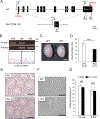
Phenotypic analysis of Fbxw5 knockout male mice. (A) Genomic structure and knockout strategy of mouse Fbxw5. Two gRNAs (red arrowheads) were designed to target the 5’-UTR region (white boxes) in exon 2 and the coding region (black boxes) of exon 9. Two primer sets (black arrows, Fw1 and Rv1; Fw2 and Rv2) were designed for gPCR. (B) Knockout and wild-type alleles were detected by gPCR using a pair of Fw1 and Rv1 and a pair of Fw2 and Rv2, respectively. Deletion of knockout allele was confirmed by Sanger sequencing. KO, He, and WT show homozygous knockout, heterozygous knockout, and wild-type mice, respectively. (C) Testis appearance of WT and Fbxw5 KO mice. Scale bar, 2 mm. (D) Bar graphs show the average testicular weight of WT and Fbxw5 KO mice. NS, no significant difference (P = 0.137). n, number of male mice in each. (E) Histological analysis of testes in WT and Fbxw5 KO mice. Scale bar, 100 μm. (F) Morphology of spermatozoa collected from the cauda epididymides in WT and Fbxw5 KO mice. Scale bar, 50 μm. (G) Bar graphs show percentages of motile spermatozoa in WT and Fbxw5 KO mice. Sperm motility was analyzed at 10 min and 2 hrs after incubation in TYH medium. NS, no significant difference (P = 0.137 for 10 min, 0.538 for 2 hrs).
Fig. 4.
Fig. 4.
Phenotypic analysis of Gm17266 knockout male mice. (A) Genomic structure and knockout strategy of mouse Gm17266. Two gRNAs (red arrowheads) were designed to target the coding region (black boxes) of exons 1 and 11. Two primer sets (black arrows, Fw1 and Rv1; Fw2 and Rv2) were designed for gPCR. (B) Knockout and wild-type alleles were detected by gPCR using a pair of Fw1 and Rv1 and a pair of Fw2 and Rv2, respectively. Deletion of knockout allele was confirmed by Sanger sequencing. KO, He, and WT show homozygous knockout, heterozygous knockout, and wild-type mice, respectively. (C) Testis appearance of WT and Gm17266 KO mice. Scale bar, 2 mm. (D) Bar graphs show average testicular weight of Gm17266 KO and WT mice. ***, significant difference (P < 0.001). n, number of male mice in each. (E) Histological analysis of testes in WT and Gm17266 KO mice. Scale bar, 100 μm. (F) Morphology of spermatozoa collected from the cauda epididymides in WT and Gm17266 KO mice. Scale bar, 50 μm. (G) Bar graphs show percentages of motile spermatozoa in WT and Gm17266 KO mice. Sperm motility was analyzed at 10 min and 2 hrs after incubation in TYH medium. NS, no significant difference (P = 0.275 for 10 min, 0.386 for 2 hrs).

Similar articles

References

    1. Abbasi F, Miyata H, Ikawa M. 2017. Revolutionizing male fertility factor research in mice by using the genome editing tool CRISPR/Cas9. Reprod. Med. Biol 17 (1), 3–10. - PMC - PubMed
    1. Abbasi F, Miyata H, Shimada K, Morohoshi A, Nozawa K, Matsumura T, Xu Z, Pratiwi P, Ikawa M 2018. RSPH6A is required for sperm flagellum formation and male fertility in mice. J. Cell Sci 131 (19), jcs221648. - PMC - PubMed
    1. Aoki Y, Tsujimura A, Kaseda K, Okabe M, Tokuhiro K, Ohta T, O’Bryan MK, Okuda H, Kitamura K, Ogawa Y, Fujiki T, Wada M, Horie S, Nishimune Y, Tanaka H 2020. Haprin-deficient spermatozoa are incapable of in vitro fertilization. Mol. Reprod. Dev 87 (5), 534–541. - PubMed
    1. Bono H, Hino K, Ui-Tei K, Naito Y 2014. CRISPRdirect: software for designing CRISPR/Cas guide RNA with reduced off-target sites. Bioinformatics 31,1120–1123. - PMC - PubMed
    1. Fang Y, Liu D, Yang Y, Zhang H, Wu H, Jiang H, Zhao L, Zhang Z 2020. Altered gene expression profiles of testicular tissues from azoospermic patients with maturation arrest. Andrologia 52 (11), e13812. - PubMed

LinkOut - more resources