. 2024 May 9;187(10):2521-2535.e21.
doi: 10.1016/j.cell.2024.04.003. Epub 2024 May 1.

RNA aggregates harness the danger response for potent cancer immunotherapy

Affiliations

RNA aggregates harness the danger response for potent cancer immunotherapy

Hector R Mendez-Gomez et al. Cell. .

Abstract

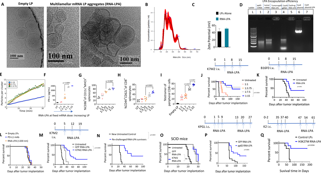
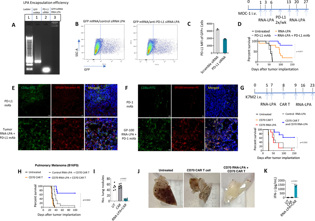
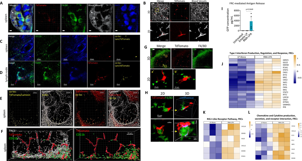
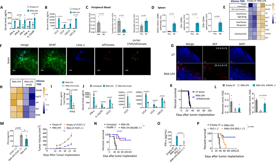
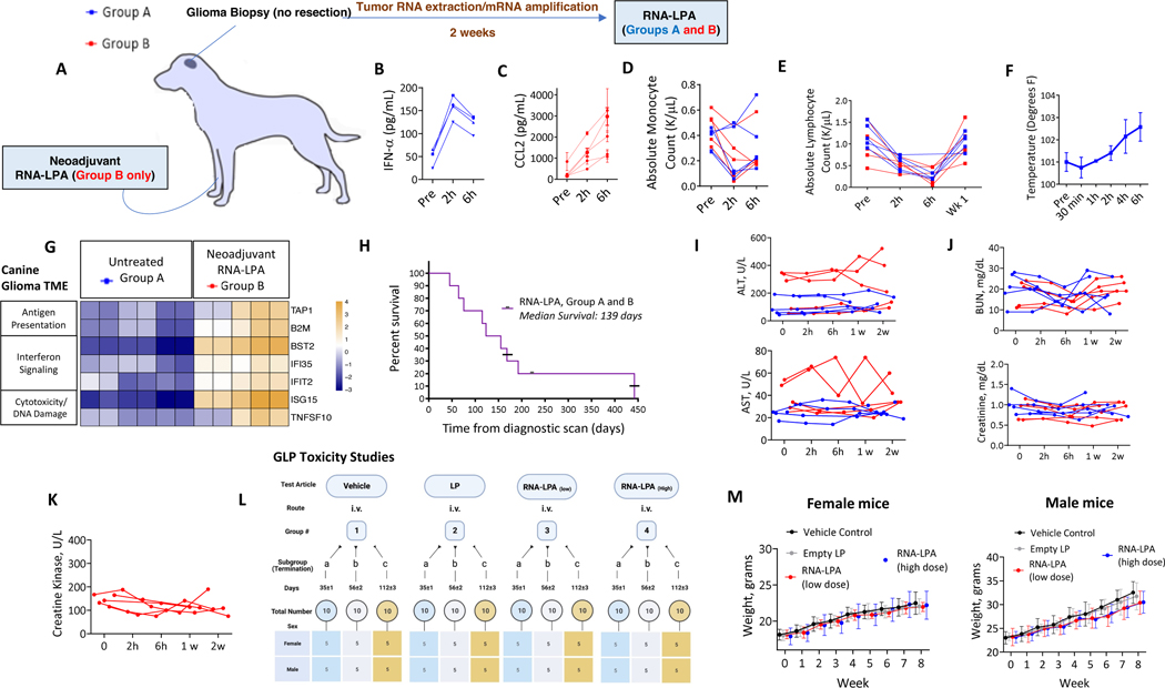
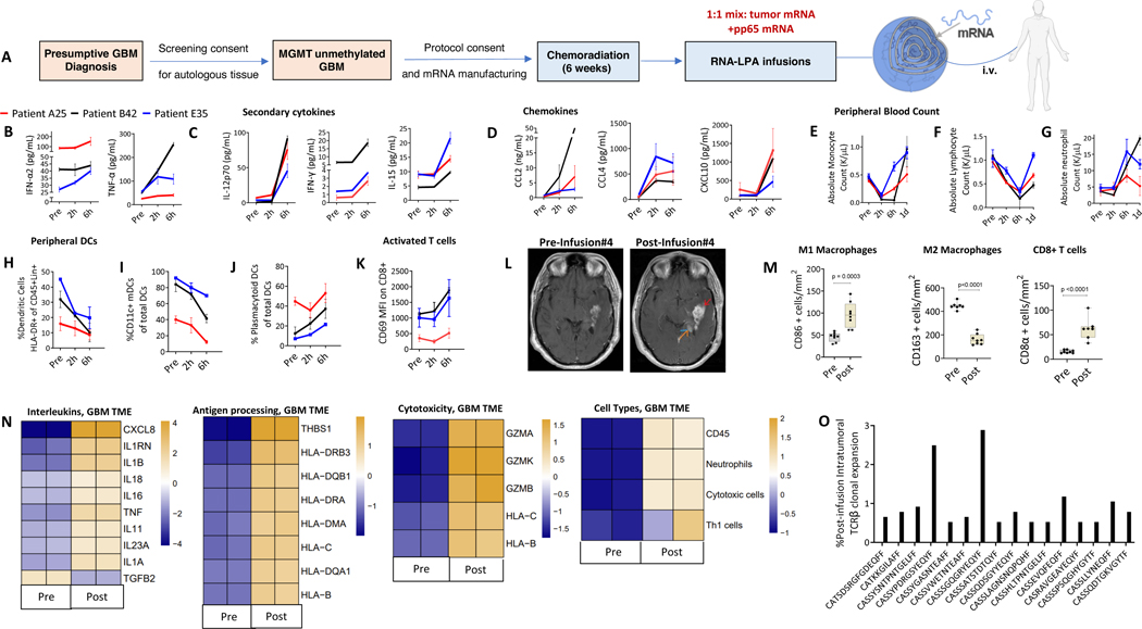
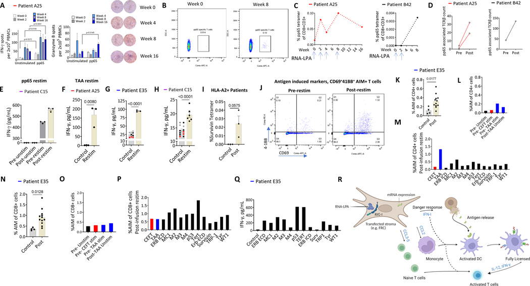
Cancer immunotherapy remains limited by poor antigenicity and a regulatory tumor microenvironment (TME). Here, we create "onion-like" multi-lamellar RNA lipid particle aggregates (LPAs) to substantially enhance the payload packaging and immunogenicity of tumor mRNA antigens. Unlike current mRNA vaccine designs that rely on payload packaging into nanoparticle cores for Toll-like receptor engagement in immune cells, systemically administered RNA-LPAs activate RIG-I in stromal cells, eliciting massive cytokine/chemokine response and dendritic cell/lymphocyte trafficking that provokes cancer immunogenicity and mediates rejection of both early- and late-stage murine tumor models. In client-owned canines with terminal gliomas, RNA-LPAs improved survivorship and reprogrammed the TME, which became "hot" within days of a single infusion. In a first-in-human trial, RNA-LPAs elicited rapid cytokine/chemokine release, immune activation/trafficking, tissue-confirmed pseudoprogression, and glioma-specific immune responses in glioblastoma patients. These data support RNA-LPAs as a new technology that simultaneously reprograms the TME while eliciting rapid and enduring cancer immunotherapy.

Keywords: brain tumors; cancer immunotherapy; cancer vaccines; glioblastoma; lipid nanoparticles; mRNA; personalized therapy; translational therapeutics.


Conflict of interest statement

Declaration of interests D.A.M. holds ownership interest in iOncologi, Inc. E.J.S is a paid consultant for Siren Biotechnology. The manuscript discusses patented technologies from H.R.M.-G., P.C., S.Q., J.M., A.P.G., J.H., W.G.S., M.R., D.A.M., and E.J.S. Patented technologies are under option to license by iOncologi, Inc.

Figures

Figure 1:
Figure 1:. RNA-LPAs optimize packaging of multiple payloads for induction of anti-tumor immunity.
A, B, C, D, Cryo-electron micrographs (a), Nanosight size distributions (b), zeta potential measurements (c), and encapsulation efficiency (d) of RNA-LPA at 1:15 mass ratios. E, RNA-LPA cluster growth over time at increasing LP:RNA ratios. F, Serum IFN-α measurements from Balb/c mice (n= 3) within 6h of i.v. RNA-LPA loaded with whole tumor derived mRNA from K7M2 (upper limit of detection for assay is 2,000 pg/mL). G, H, Activated DCs (g) and central memory CD8 T cells (h) from spleens of Balb/c mice (n= 3–5/group) bearing K7M2 pulmonary sarcomas harvested 24h after a third weekly dose of i.v. RNA-LPA loaded with tumor derived mRNA (from K7M2). I, C57Bl/6 mice (n=7–8/group) were implanted with B16F10-OVA subcutaneously on day 0 and injected with OT-1 cells intravenously on the next day. Tetramer+ T cells from spleens were assessed after third weekly dose of i.v. OVA specific RNA-LPA. J, Survival curve of Balb/c mice (n= 8/group) bearing pulmonary K7M2 sarcomas treated with i.v. tumor derived RNA-LPA. K, Survival curve of C57Bl/6 mice (n=8) bearing pulmonary B16F0 melanomas treated i.v. with B16F0 tumor derived RNA-LPAs. L, Survival curve of C57Bl/6 mice (n= 8/group) bearing pulmonary B16F0 melanomas treated with empty LPs, PD-L1 mAbs or i.v. tumor derived RNA-LPA filtered for aggregates < 200 nm. M, Survival curve of Balb/c mice (n= 5–8/group) bearing pulmonary K7M2 sarcomas treated with three weekly i.v. GFP or K7M2 tumor derived RNA-LPA (data is aggregate of two separate experiments). N, Re-challenge of long-term surviving Balb/c mice (n=7–8/group) previously treated with K7M2 tumor derived RNA-LPA inoculated at 120 days with K7M2 i.v. versus a new cohort of untreated mice. O, Survival curve of SCID Fox-Chase mice on Balb/c background (n= 7–8/group) bearing pulmonary K7M2 sarcomas treated with three weekly i.v. GFP or K7M2 tumor derived RNA-LPA. P, Survival curve of C57Bl/6 mice (n = 14) bearing intracranial KR158b gliomas transduced with pp65 and treated i.v. with weekly GFP or pp65 RNA-LPAs. pp65 mRNA constructs made with pseudouridines. Q, Survival curve of C57Bl/6 mice (n= 7–8/group) implanted neonatally with K2 midline gliomas expressing H3K27M and treated with i.v. H3K27M encoding RNA-LPA. Significance was determined via parametric student’s t-test (F-I), and log-rank test (J-Q). Error bars are reported as the standard error of the mean (F) and standard deviation of the mean (C, G-I).
Figure 2:
Figure 2:. RNA-LPAs co-deliver multiple payloads and sensitize response to synergistic immunotherapy.
A, Encapsulation efficiency of RNA-LPA simultaneously loaded with GFP mRNA and anti-PD-L1 siRNA. B, FACS plots of RNA-LPA transfected DC2.4 cells. C, GFP transfected cells from (B) were assessed for PD-L1 expression by flow cytometric analysis of mean fluorescent intensity (MFI). D, In C57Bl/6 mice bearing MOC-1 tumors (n=10/group), i.v. RNA-LPAs were administered in combination with anti-PD-L1 mAbs given as a loading dose and maintained twice weekly until last RNA-LPA treatment. Data censored at 150 days. E, C57Bl/6 mice (n=8/group) bearing pulmonary B16F0 melanomas treated with anti-PD-L1 mAbs, with or without B16F0 tumor derived RNA-LPAs i.v., before established tumors/lungs were harvested for IHC of tumor infiltrating lymphocytes specific for gp-100. F, C57Bl/6 mice (n=8/group) bearing pulmonary B16F0 melanomas were treated i.v. with P-mel splenocytes (1x10⁷ /mouse) and PD-1 mAb with or without gp-100 RNA-LPA i.v. before established tumors/lungs were harvested for IHC of tumor infiltrating lymphocytes specific for gp-100. G, In Balb/c mice bearing K7M2 sarcomas (n= 8/group), RNA-LPAs (i.v.) began on day 5 after tumor tail vein inoculation, day 7 (~24h before CAR T administration) and weekly thereafter (x3). H, In C57Bl/6 mice bearing B16F0 (n = 12/group), RNA-LPAs (i.v.) began on day 1 after tumor tail-vein inoculation, day 4 (~24 h before CAR T administration) and weekly thereafter (x3). I, J, In C57Bl/6/c mice bearing B16F0 (n= 6/group) treated with CD70 RNA-LPA and CD70 CAR T as above, CLARITY imaging of lung nodules was performed to assess tumor burden across treatment groups. K, Following CD70 RNA-LPA and CD70 CAR T cells, splenocytes were harvested for CAR T enumeration and functional assessment in culture with transduced (CD70pos) K7M2 tumors. Significance was determined via and log-rank test (D, G, H), Mann-Whitney test (I) and parametric student’s t-test (K). Error bars are reported as standard deviation (C) and standard error of the mean (I, K). Scale bars: E, F, 10 μm.
Figure 3:
Figure 3:. RNA-LPAs transfect lymphoreticular stroma eliciting mRNA expression, antigen release and danger response.
A, Cross section of the whole spleen 2 days following i.v. injection with Cre RNA-LPA in Ai14 tdTomato reporter mice. Yellow dashed box maps to magnified panels ‘G.’ B, 3D and magnified 2D image of vasculature in splenic white pulp demonstrating that tdTomato+ cells are perivascular. C, D, Voxel based co-localization analysis in spleen and lymph nodes of tdTomato with FRC markers (i.e. laminin) following Cre RNA-LPAs (i.v.). E, 3D surface-based co-localization between FRCs (laminin+) with tdTomato. Colocalization is displayed as an independent channel in yellow. F, tdTomato+ FRCs in close contact with CD11b+ cells. G, Top row maps to yellow box in panel 'A' with 3D overlay of tdTomato with F4/80; bottom row displays two inlaid boxes at higher magnification in 2D. H, Left column shows 2 different tdTomato cells (in 2D) interfacing with macrophages (separate from ‘G’); right column, 3D capture of these cells, rotating to display maximum contact ‘confluence’ points identified via voxel co-localization (blue). I, GFP ELISA from supernatants (n=3–4/group) of human in vitro FRCs transfected with GFP RNA-LPA. Significance determined via parametric student’s t-test. J, K, L, Significantly upregulated gene sets by RNA sequencing for human in vitro FRCs transfected with RNA-LPA (n=3, technical replicates) compared to Empty LP treated cells (n=3, technical replicates). Heatmaps of differential gene expression showing fold change in gene expression, with yellow indicating upregulation and blue indicating downregulation: Type I Interferon Production, Regulation, and Response (J), RIG-I-Like Receptor Pathway (K), Chemokine and Cytokine production, secretion, and receptor interaction (L). Scale bars: A, 200 μm; B top, C, D, F, G top, 50 μm; B bottom, G bottom, H left, 10 μm; E, 150 μm; H right, 5 μm.
Figure 4:
Figure 4:. RNA-LPAs chemoattract PBMCs, reprogram the TME and mediate activity through stimulation of intracellular PRRs.
A, B, Cytokine/chemokine response panel from C57Bl/6 mice (n=3/group) 6 hours post treatment with RNA-LPAs i.v. C, Absolute counts of peripheral white blood cells from C57Bl/6 mice (n=5/group) 6h after i.v. luciferase RNA-LPAs. D, Absolute counts of activated DCs and activated T cells in spleens of C57Bl/6 mice (n=5/group) harvested 6h after i.v. luciferase RNA-LPAs. E, RNA sequencing of established KR158b intracranial tumors harvested 48h after a single tumor derived RNA-LPA. F, Co-localization analysis in brains from K2 glioma bearing animals with LYVE-1 and tdTomato following Cre RNA-LPAs (i.v.) in Ai14 tdTomato reporter mice. G, Dual Reporter mice (CCR2/CX3CR1) vaccinated 7 days following KR158-luc tumor implantation. Tissues were collected 48 hours after vaccination and assessed for % infiltrating RFP+ leukocytes (in total cell count, quantified by DAPI). Values are combined n=2 for untreated and total tumor mRNA-LPA vaccinated mice. H, RNA sequencing of established KR158b intracranial tumors harvested 48h after a single tumor derived RNA-LPA with either silenced (modRNA) or unsilenced RNA payloads. I, J, Serum cytokine/chemokine response panel from C57Bl/6 mice (n=3/group) 6h post treatment with silenced or unsilenced i.v. RNA-LPA. K, Survival curve of C57Bl/6 mice (n=12–14/group) bearing pulmonary B16F0 melanomas treated i.v. with a total of six RNA-LPAs concomitantly with IFNAR1 mAbs. L, Absolute monocyte and lymphocyte count in peripheral blood of animals within 24h of RNA-LPA treatment in wild-type and IFNAR1 knock-out animals (n=3–4/group). M, Serum analysis of IFN-α (left), n=3/group and tumor volume growth curve (right) from C57Bl/6 wild-type versus TLR7 knock-out mice (n=5–8/group) bearing subcutaneous B16F10-OVA melanomas treated i.v. with OVA RNA-LPA. N, Survival curve from C57Bl/6 wild-type versus MYD88 knock-out mice (n= 7–8/group) bearing pulmonary B16F10-OVA melanomas treated i.v. with OVA RNA-LPA. O, P, Serum analysis of IFN-α (O), n=3/group, and survival curve (P) from C57Bl/6 wild-type versus RIG-I knock-out mice (n=7–8/group) bearing pulmonary B16F10-OVA melanomas treated i.v. with OVA RNA-LPA. Significance determined via parametric student’s t-test (A, B, C, D, I, J, L, M (left), O), mixed effects analysis/ANOVA (M, right) and log-rank test (K, N, P). Error bars reported as the standard error of the mean (A, B, C, D, I, J, M (left), O) and the standard deviation of the mean (M (left), O). Scale bars: F, 50 μm; G, 200 μm.
Figure 5:
Figure 5:. RNA-LPAs mobilize PBMCs and reprogram the TME of client-owned canines with terminal gliomas.
A, Study schema of client-owned canines with terminal gliomas enrolled to receive RNA-LPA. Group A: Adjuvant tumor derived RNA-LPA alone” vs. “Group B: Neoadjuvant pp65-RNA-LPA followed by tumor derived RNA-LPA. Tumor derived mRNA was generated after whole tumor RNA extraction from canine glioma samples followed by cDNA library generation by RT-PCR and mRNA amplification through in vitro transcription reaction. B, C, Serum from canines was obtained for analysis of IFN-α (Group A, n=4) (B) and CCL2 (Group B, n=5) (C). D, E, Absolute peripheral blood monocyte (D) and lymphocyte (E) count from canines (Group A and B, n= 10) pre, 2, and 6h post-initial RNA-LPA. F, Temperature measurements from first 4 Group A canines following RNA-LPA (Subject 1–2, after initial infusion; Subject 3–4, after 3rd infusion). G, Nanostring analysis of untreated canine glioma specimens (Group A, n=3, in duplicate) or within 48h of RNA-LPA (Group B, n=3, 2 in duplicate, 1 singlet). H, Survival curve of canines (Group A and B, n= 10) bearing intracranial gliomas undergoing treatment with RNA-LPAs. Crosses at 168 days and 442 days delineate 2 canines receiving additional therapies: pre-vaccine CCNU (168 days) and post-vaccine radiation therapy (442 days). I, J, K, Liver (I) and renal (J) function tests from serum enzymes in Group A (blue) and B (red) subjects and creatinine kinase (K) from Group B subjects following RNA-LPA administrations. L, GLP toxicology study groups. Treatment groups consisted of Empty LPs or LPAs of tumor mRNA (amplified from KR158b) and pp65 mRNA mixed 1:1. M, Weights from GLP toxicology studies in animals undergoing treatment with RNA-LPAs. Error bars reported as standard error of the mean (C, F) from individual subject replicates (C) and pooled replicates across canines (F). Error bars reported as standard deviation in panel (M).
Figure 6:
Figure 6:. RNA-LPAs mediate rapid cytokine/chemokine release, mobilization of PBMCs, and TME remodeling in human glioblastoma patients.
A, Study schema of MGMT unmethylated glioblastoma patients enrolled to receive RNA-LPA. B, C, D, Cytokine/chemokine sera measurements pre, 2, and 6h post-RNA-LPA. E, F, G, Absolute counts of peripheral blood monocytes (E), lymphocytes (F), and neutrophils (G). H, I, J, Fraction of APCs (H) and DC subtypes including mDCs (I) and plasmacytoid DCs (pDCs) (J). K, Mean fluorescent intensity (MFI) of activated CD8 T cells. L, T1 axial MRI images pre-infusion#4 and increasing enhancement 1-month post-infusion. M, Box-plot of intratumoral M1/M2 macrophages and CD8 T cells following IHC preparation and staining. Error bars represent min to max values and p values determined by unpaired t test. N, Nanostring analysis of pre and post-infusion tumor specimens in duplicate. O, Selected TCRβ expanded clonotypes (from zero, pre-infusion) greater than 0.5% in the TME of post-infusion tumor sample. Error bars are reported as standard error of the mean (B, C, D, E, F, G, H, I, J, K).
Figure 7:
Figure 7:. RNA-LPAs induce antigen specific T cell responses in human glioblastoma patients.
A, IFN-γ and granzyme B ELISpots using unstimulated and pp65 re-stimulated PBMCs from Patient A25. B, C, Flow cytometric analysis of antigen specific T cells in Patients A25 (B, C) and B42 (C) by tetramer staining for HLA-A2 restricted pp65 epitope. Blue arrows indicate the timing of vaccine doses. D, Expanded pp65-specific TCRβ counts in Patient A25 and B42 after 2 and 4 RNA-LPA infusions, respectively. E, PBMCs from Patient C15 cultured with overlapping pp65 peptide pool. F, PBMC replicates from patient A25 in triplicate co-cultured with overlapping TAA peptide pool (n=12) for glioma associated antigens with supernatants harvested after 48h for IFN-γ response versus pre-infusion-TAA stimulated control samples. G, PBMCs from Patient E35 co-cultured with overlapping peptide pool for glioma associated antigens (n=12) with supernatants harvested after 9-day culture for IFN-γ response versus control samples (Red Dots: within 2h of initial infusion, unstimulated; Grey: within 2h of initial infusion, TAA stimulated; Black: post-infusions, unstimulated). H, PBMCs from Patient C15 co-cultured with overlapping peptide pool for glioma associated antigens (n=12) with supernatants harvested after 9-day culture for IFN-γ response versus control samples (Red Dots: pre-infusion, unstimulated; Grey: pre-infusion, TAA stimulated; Black: post- 3rd and 4th infusions, unstimulated). I, PBMCs from the 3 HLA-A2+ patients stained for Survivin tetramer in post-infusion TAA stimulated samples compared with post-infusion TAA unstimulated controls. J, FACS plots demonstrating increase in double positive antigen specific CD3+ CD4+ T cells (CD69+, 4–1BB+) in post-vaccinated patient after culture with overlapping peptides from 12 TAAs. K, L, M, Percentage of antigen specific CD4 cells from Patient E35 quantified via AIM assay after in vitro co-culture with overlapping peptide from 12 glioma associated antigens. Each dot from post-vaccinated samples denotes antigen specific T cells in response to one overlapping peptide (K) versus control samples shown in (L). Response to each distinct antigen (black bars) is shown in (M) versus whole TAA mix (all 12 peptides, blue bar) and versus positive control with CEFT (red bar). N, O, P, Percentage of antigen specific CD8 cells from Patient E35 quantified via AIM assay after in vitro co-culture with overlapping peptide from 12 glioma associated antigens. Each dot from post-vaccinated samples denotes antigen specific T cells in response to one overlapping peptide (N) versus control samples shown in (O). Response to each distinct antigen (black bars) is shown in (P) versus whole TAA mix (all 12 peptides, blue bar) and versus positive control with CEFT (red bar). Q, IFN-γ recall response from Patient E35 following single antigen re-stimulation. R, Proposed mechanism for RNA-LPA mediated immunity (image created with Biorender.com). P values determined via parametric student’s t-test (A, F, G, H, K, N). Error bars reported as the standard error of the mean (A, E, F, G, H, I, K, N).

Update of

  • mRNA aggregates harness danger response for potent cancer immunotherapy.
    Mendez-Gomez HR, DeVries A, Castillo P, Stover BD, Qdaisat S, Von Roemeling C, Ogando-Rivas E, Weidert F, McGuiness J, Zhang D, Chung MC, Li D, Zhang C, Marconi C, Campaneria Y, Chardon-Robles J, Grippin A, Karachi A, Thomas N, Huang J, Milner R, Sahay B, Sawyer WG, Ligon JA, Silver N, Simon E, Cleaver B, Wynne K, Hodik M, Molinaro A, Guan J, Kellish P, Doty A, Lee JH, Carrera-Justiz S, Rahman M, Gatica S, Mueller S, Prados M, Ghiaseddin A, Mitchell DA, Sayour EJ. Mendez-Gomez HR, et al. medRxiv [Preprint]. 2023 Mar 17:2023.03.12.23287108. doi: 10.1101/2023.03.12.23287108. medRxiv. 2023. Update in: Cell. 2024 May 9;187(10):2521-2535.e21. doi: 10.1016/j.cell.2024.04.003. PMID: 36993772 Free PMC article. Updated. Preprint.

Comment in

  • RNA delivery heats up cold tumours.
    Villanueva MT. Villanueva MT. Nat Rev Drug Discov. 2024 Jul;23(7):497. doi: 10.1038/d41573-024-00098-0. Nat Rev Drug Discov. 2024. PMID: 38858569 No abstract available.

Similar articles

Cited by

References

    1. Kranz LM, Diken M, Haas H, Kreiter S, Loquai C, Reuter KC, Meng M, Fritz D, Vascotto F, Hefesha H, et al. (2016). Systemic RNA delivery to dendritic cells exploits antiviral defence for cancer immunotherapy. Nature. 10.1038/nature18300. - DOI - PubMed
    1. Kreiter S., Selmi A., Diken M., Koslowski M., Britten CM., Huber C., Tureci O., and Sahin U. (2010). Intranodal vaccination with naked antigen-encoding RNA elicits potent prophylactic and therapeutic antitumoral immunity. Cancer research 70, 9031–9040. 10.1158/0008-5472.CAN-10-0699. - DOI - PubMed
    1. Sahin U, Oehm P, Derhovanessian E, Jabulowsky RA, Vormehr M, Gold M, Maurus D, Schwarck-Kokarakis D, Kuhn AN, Omokoko T, et al. (2020). An RNA vaccine drives immunity in checkpoint-inhibitor-treated melanoma. Nature 585, 107–112. 10.1038/s41586-020-2537-9. - DOI - PubMed
    1. Grabbe S, Haas H, Diken M, Kranz LM, Langguth P, and Sahin U. (2016). Translating nanoparticulate-personalized cancer vaccines into clinical applications: case study with RNA-lipoplexes for the treatment of melanoma. Nanomedicine (Lond) 11, 2723–2734. 10.2217/nnm-2016-0275. - DOI - PubMed
    1. Holtkamp S, Kreiter S, Selmi A, Simon P, Koslowski M, Huber C, Tureci O, and Sahin U. (2006). Modification of antigen-encoding RNA increases stability, translational efficacy, and T-cell stimulatory capacity of dendritic cells. Blood 108, 4009–4017. 10.1182/blood-2006-04-015024. - DOI - PubMed

Publication types

MeSH terms

LinkOut - more resources