Review
. 2024 May 15;112(10):1568-1594.
doi: 10.1016/j.neuron.2024.04.011. Epub 2024 May 1.

Consciousness and sleep

Affiliations
Review

Consciousness and sleep

Giulio Tononi et al. Neuron. .

Abstract

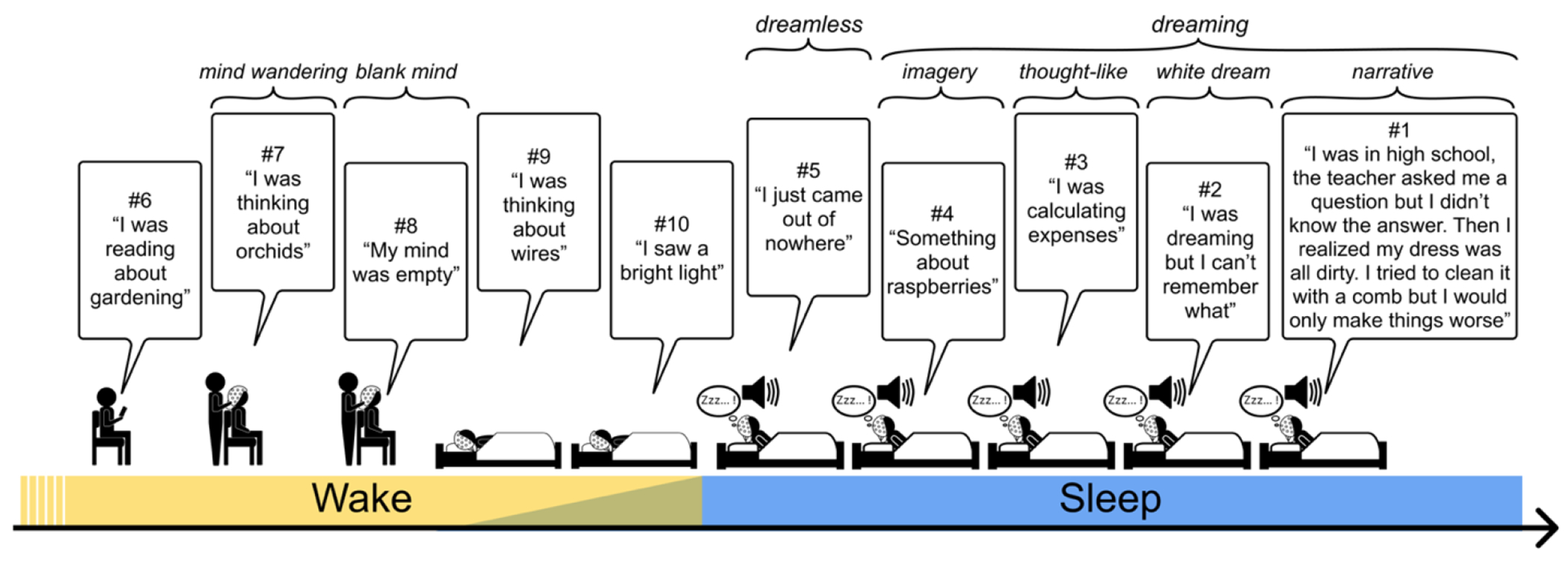
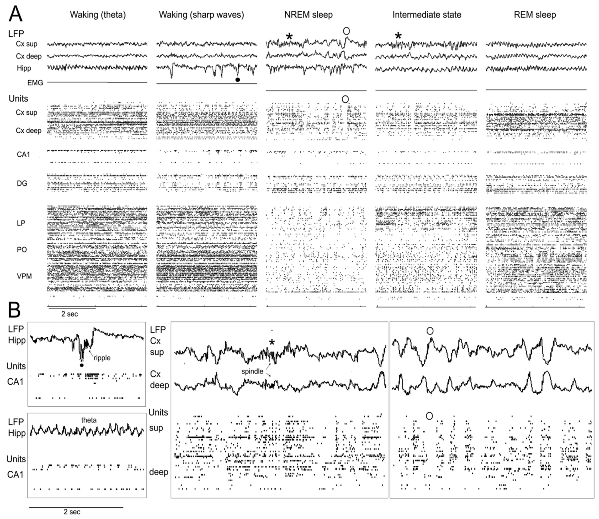
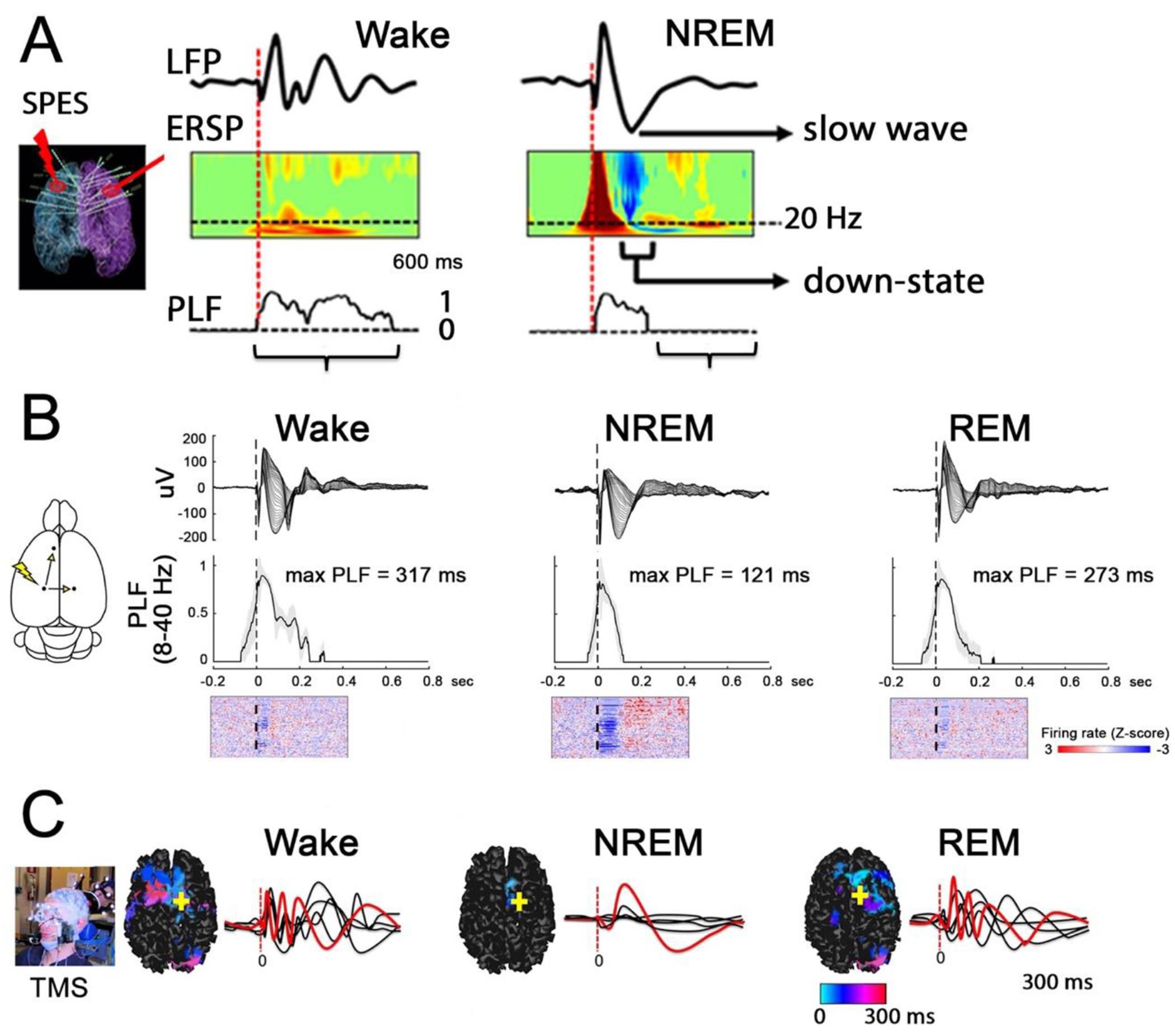
Sleep is a universal, essential biological process. It is also an invaluable window on consciousness. It tells us that consciousness can be lost but also that it can be regained, in all its richness, when we are disconnected from the environment and unable to reflect. By considering the neurophysiological differences between dreaming and dreamless sleep, we can learn about the substrate of consciousness and understand why it vanishes. We also learn that the ongoing state of the substrate of consciousness determines the way each experience feels regardless of how it is triggered-endogenously or exogenously. Dreaming consciousness is also a window on sleep and its functions. Dreams tell us that the sleeping brain is remarkably lively, recombining intrinsic activation patterns from a vast repertoire, freed from the requirements of ongoing behavior and cognitive control.

Keywords: bistability; disconnection; dreaming; neural correlates; perception.


Conflict of interest statement

Declaration of interests G.T. holds an executive position and has a financial interest in Intrinsic Powers, Inc., a company whose purpose is to develop a device that can be used in the clinic to assess the presence of consciousness in patients. G.T. also holds a patent concerning such a device.

Figures

Figure 1:
Figure 1:. Experience sampling in wakefulness and sleep.
Examples of experience sampling at various times during the day, at sleep onset, and during sleep (serial awakening paradigm). The examples also introduce the nomenclature used throughout the review.
Figure 2.
Figure 2.. Neural activity in sleep and wakefulness.
A. Recording of local field potentials (LFP) and unit activity (units) in a freely moving rat during wakefulness and sleep, using a high density Neuropixels probe (384 sites) spanning, from dorsal to ventral, parietal association cortex (all layers), dorsal hippocampus (CA1 and dentate gyrus, DG), and multiple thalamic nuclei (lateral posterior/pulvinar, LP; posterior, Po; ventroposterior medial, VPM). Three LFP channels are shown at the top (superficial and deep cortex, CA1, stratum radiatum) for each behavioral state. EMG: electromyogram. Five behavioral states are shown, from left to right: active wakefulness with strong theta activity evident especially in the hippocampal LFP; quiet wakefulness with several hippocampal sharp waves (one indicated by a black filled circle); NREM sleep with a spindle (asterisk) and several slow waves (one indicated by the open circle) in the cortical LFPs and hippocampal sharp waves; intermediate state (transition from NREM sleep to REM sleep) with dense spindling in cortex and theta activity in the hippocampus; REM sleep with strong theta activity in both cortex and hippocampus. B. Major rhythms and oscillations are shown at higher magnification: sharp wave coupled to a ripple in the hippocampal LFP, associated with strong CA1 firing; hippocampal theta activity with rhythmic unit firing in CA1 and DG; spindle detected in superficial and deep cortical layer, and associated cortical firing; many cortical slow waves, one indicated by the open circle. Note the inverted polarity in superficial and deep layers, and the association with OFF periods (100-200 milliseconds of unit silence).
Figure 3:
Figure 3:. Neural correlates of loss of consciousness in sleep.
(A) Significant increase in slow wave activity (1-4 Hz) in subjects reporting no experiences vs. dreaming upon awakening within NREM sleep (in yellow) and within REM sleep (in orange). A conjunction analysis is shown in brown. The results are based on high-density EEG data reported in Siclari et al., 2017. The increase in slow wave activity during dreamless sleep is localized primarily over posterior-central cortical regions. LL (left lateral view), LM (left medial), RL (right lateral), RM (right medial). (B). Increased high-frequency activity (20-50 Hz) when subjects reported dreaming of faces, vs. dreaming of other contents, just before awakening from REM sleep. The increase in high-frequency activity is localized over the before awakening from REM sleep. The increase in high-frequency activity is localized over the fusiform gyrus and is maximal a few seconds before the awakening (from Siclari et al., 2017).
Figure 4:
Figure 4:. Neural mechanisms of loss of consciousness in dreamless sleep.
A. During wakefulness, single-pulse electrical stimulation (SPES) triggers a complex, prolonged response in the local field potential (LFP) recorded with simultaneous stereotactic EEG; This is also evident in the event-related spectral perturbation (ERSP); moreover, the phase-locking factor (PLF) is significantly elevated for more than 500 milliseconds, indicating sustained causal interactions triggered by the stimulus. During deep NREM sleep, SPES triggers an LFP response resembling a slow wave, associated with the suppression of high frequencies in the ESRP, and followed by the abrupt decay of the PLF (adapted from Pigorini et al., 2015). B. Schematic of the rat brain displaying the location of stimulating and recording probes in one rat (left) and examples of event related potentials (top), max phase locking factor in the 8-40 Hz range (middle) and single-unit peri-stimulus time histograms in a parietal probe locked to the onset of the electrical stimulation (bottom) across behavioral states in one rat. Units are sorted by depth. The vertical dashed line indicates the onset of the electrical stimulation (adapted from Cavelli et al., 2023). C. Spatiotemporal cortical current maps of TMS-induced activity during wakefulness, NREM, and REM sleep in a representative subject. The inset is the setup for TMS/EEG. For each significant time sample, maximum current sources are plotted and color-coded on the subject’s MRI according to the activation latency (light blue, 0 milliseconds; red, 300 milliseconds). The yellow cross marks the TMS target on the cortical surface. To the right is the evoked response, in red for an EEG channel near the stimulation site, and in black for four representative channels further away (adapted from Massimini et al., 2010).
Figure 5:
Figure 5:. Comparison of brain activity between REM sleep and resting wakefulness.
Cortical areas more and less active during REM sleep compared to quiet wakefulness are plotted in red and dark blue, respectively. Significant clusters were obtained by (Fox et al., 2013) using an activation likelihood estimation analysis across 6 PET studies (Maquet et al., 1996; Nozfinger et al., 1997; Braun et al., 1997; Braun et al., 1998; Maquet et al., 2000; Peigneux et al., 2001) for a total of 61 subjects. The figure was created using a standard template (BrainNet Viewer (http://www.nitrc.org/projects/bnv/, Xia et al., 2013). Node locations centered on peak activation / deactivation coordinates as reported by (Fox et al., 2013), then projected to cortical surface with node sizes proportional to the number of voxels for each cluster. Subcortical structures that were more (pons, midbrain and caudate nucleus) or less activated (superior longitudinal fasciculus) during REM sleep than during wakefulness according to the (Fox et al., 2013) meta-analysis are not displayed in the figure.

References

    1. Freud S, and Strachey J (2010). The interpretation of dreams (Basic Books).
    1. Tononi G, Boly M, Massimini M, and Koch C (2016). Integrated information theory: from consciousness to its physical substrate. Nat Rev Neurosci 17, 450–461. 10.1038/nm.2016.44. - DOI - PubMed
    1. Cirelli C, and Tononi G (2008). Is sleep essential? PLoS Biol 6, e216. - PMC - PubMed
    1. Noreika V, Valli K, Lahtela H, and Revonsuo A (2009). Early-night serial awakenings as a new paradigm for studies on NREM dreaming. Int J Psychophysiol 74, 14–18. 10.1016/j.ijpsycho.2009.06.002. - DOI - PubMed
    1. Siclari F, Larocque JJ, Postle BR, and Tononi G (2013). Assessing sleep consciousness within subjects using a serial awakening paradigm. Front Psychol 4, 542. 10.3389/fpsyg.2013.00542. - DOI - PMC - PubMed

LinkOut - more resources