

A pupillary contrast response in mice and humans: Neural mechanisms and visual functions
- PMID: 38697114
- PMCID: PMC11257825
- DOI: 10.1016/j.neuron.2024.04.012
A pupillary contrast response in mice and humans: Neural mechanisms and visual functions
Abstract
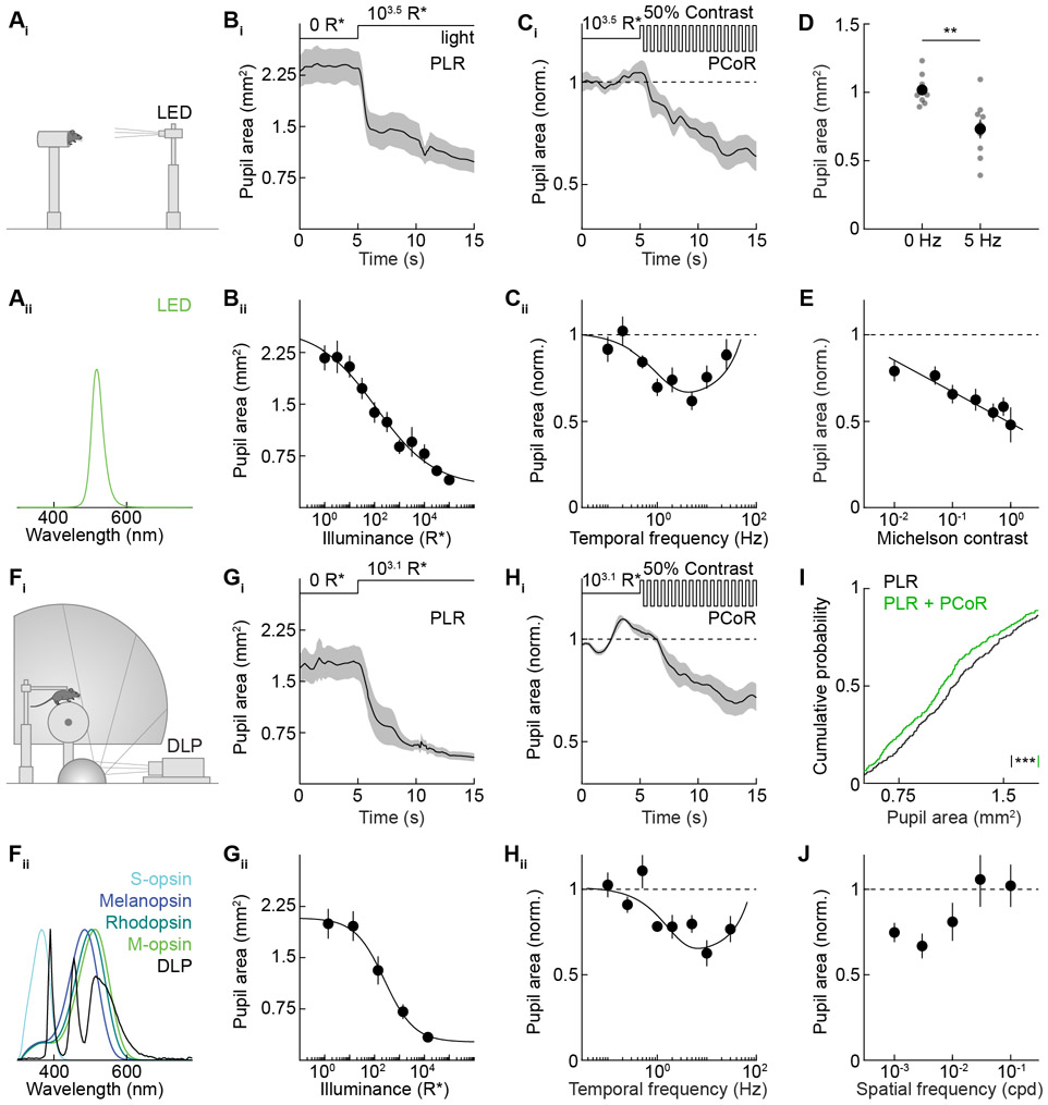
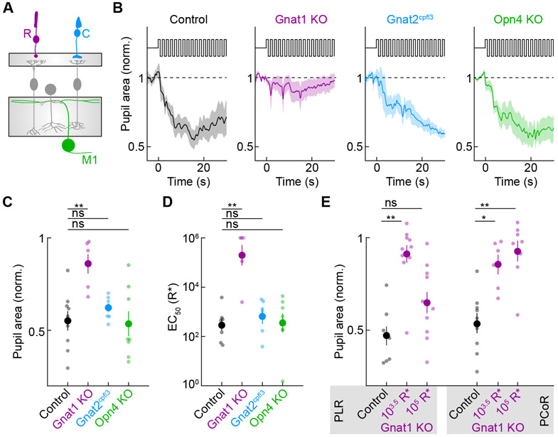
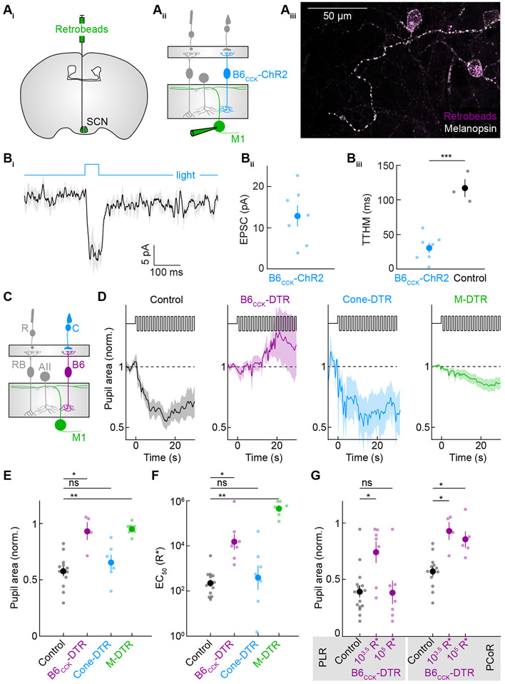
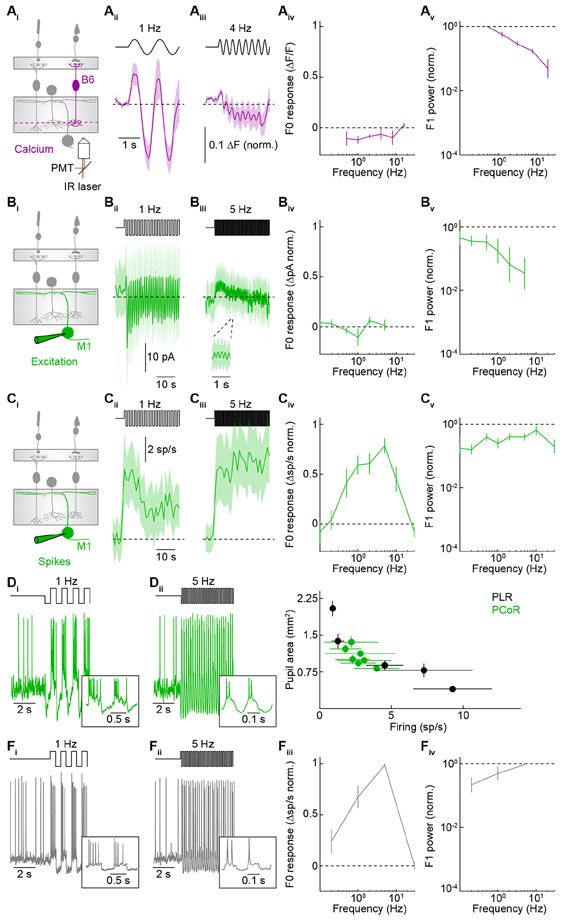
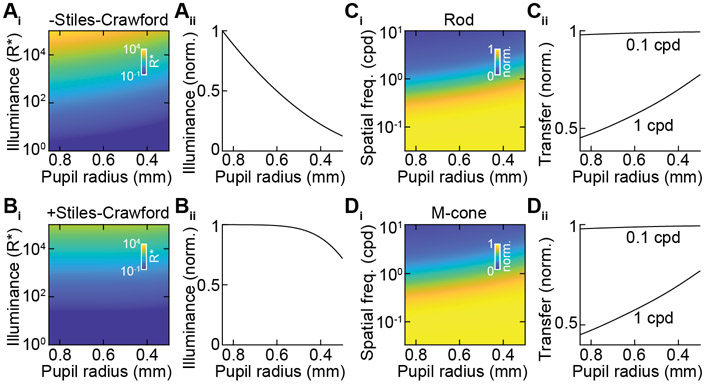
In the pupillary light response (PLR), increases in ambient light constrict the pupil to dampen increases in retinal illuminance. Here, we report that the pupillary reflex arc implements a second input-output transformation; it senses temporal contrast to enhance spatial contrast in the retinal image and increase visual acuity. The pupillary contrast response (PCoR) is driven by rod photoreceptors via type 6 bipolar cells and M1 ganglion cells. Temporal contrast is transformed into sustained pupil constriction by the M1's conversion of excitatory input into spike output. Computational modeling explains how the PCoR shapes retinal images. Pupil constriction improves acuity in gaze stabilization and predation in mice. Humans exhibit a PCoR with similar tuning properties to mice, which interacts with eye movements to optimize the statistics of the visual input for retinal encoding. Thus, we uncover a conserved component of active vision, its cell-type-specific pathway, computational mechanisms, and optical and behavioral significance.
Keywords: Stiles-Crawford effect; active vision; eye movements; ipRGCs; pupil; retina; rod pathways; visual behavior; visual optics.
Copyright © 2024 Elsevier Inc. All rights reserved.
Conflict of interest statement
Declaration of interests The authors declare no competing interests.
Figures
References
-
- Gibson JJ (2014). The ecological approach to visual perception: classic edition (Psychology press; ).
-
- Findlay JM, and Gilchrist ID (2003). Active Vision: The Psychology of Looking and Seeing (Oxford University Press; ).
-
- Yarbus AL, and Haigh B (1973). Eye Movements and Vision (Plenum Press; ).
MeSH terms
Grants and funding
LinkOut - more resources
Full Text Sources
Molecular Biology Databases
Research Materials
