. 2024 Jul 16;30(8):1041-1057.
doi: 10.1261/rna.079837.123.

A novel reporter for helicase activity in translation uncovers DDX3X interactions

Affiliations

A novel reporter for helicase activity in translation uncovers DDX3X interactions

Kevin C Wilkins et al. RNA. .

Abstract

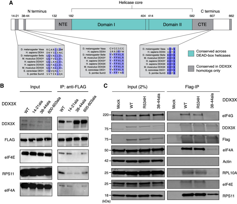
DDX3X regulates the translation of a subset of human transcripts containing complex 5' untranslated regions (5' UTRs). In this study, we developed the helicase activity reporter for translation (HART), which uses DDX3X-sensitive 5' UTRs to measure DDX3X-mediated translational activity in cells. To directly measure RNA structure in DDX3X-dependent mRNAs, we used SHAPE-MaP to determine the secondary structures present in DDX3X-sensitive 5' UTRs and then used HART to investigate how sequence alterations influence DDX3X sensitivity. Additionally, we identified residues 38-44 as potential mediators of DDX3X's interaction with the translational machinery. HART revealed that both DDX3X's association with the translational machinery and its helicase activity are required for its function in promoting the translation of DDX3X-sensitive 5' UTRs. These findings suggest DDX3X plays a crucial role in regulating translation through its interaction with the translational machinery during ribosome scanning and establish the HART reporter as a robust, lentivirally encoded, colorimetric measurement of DDX3X-dependent translation in cells.

Keywords: RNA helicases; RNA structure; reporter genes; translational control.


Figures

FIGURE 1.
FIGURE 1.
The HART uses DDX3X-sensitive 5′ UTRs to measure the translational activity of DDX3X. (A) Selection of DDX3X-sensitive 5′ UTRs for HART. Validated DDX3X-sensitive 5′ UTR from prior ribosome profiling and in vitro translation as well as negative controls were cloned into the HART reporter. (B) Diagram of HART. HART is constructed around a bidirectional promoter, with one arm directing the transcription of a control 5′ UTR and eGFP, and the other arm featuring a DDX3X-sensitive 5′ UTR followed by mCherry. An ornithine decarboxylase (ODC) degron (d4) shortens the mCherry half-life. The HART ratio (mCherry/eGFP) can be used to measure the translational activity of DDX3X. (C) Table of features for the 5′ UTRs from C. Features cataloged include length, GC content, RNA minimum free energy (MFE) prediction using the ViennaRNA Package (Lorenz et al. 2011), and the number of upstream AUGs and near-cognate codons. (D) Western blot for cells in A. HCT116 degron cells were treated with either auxin or DMSO, and then lysates were collected and resolved by SDS-PAGE and used for western blotting with antibodies against actin and DDX3X. (E) HART ratio in HCT116 degron cells analyzed with flow cytometry. HCT116 degron cells were lentivirally transduced with HART constructs with indicated 5′ UTRs upstream of mCherry. After 48 h from the addition of either DMSO or auxin, which induces degradation of endogenous DDX3X, the fluorescent signal of cells was measured by fluorescent cytometry. The HART ratio (mCherry/eGFP) was calculated for each cell and averaged across replicate wells. Data were obtained in three separate experiments for a total of nine replicates. Statistical significance was determined by unpaired t-test: (ns) P > 0.05, (*) P ≤ 0.05, (**) P ≤ 0.01, (***) P ≤ 0.001, (****) P ≤ 0.0001. (F) HART ratio in HCT116 degron cells analyzed by microscopy. HCT116 degron cells were transduced with HART construct for ODC1 or RAC1. After 48 h from the addition of either DMSO or auxin, the cells were fixed and imaged using an InCell Analyzer 6500HS. The HART ratio was calculated for each cell and averaged across replicate wells. Data were obtained in two separate experiments each for a total of 36 replicates. Statistical significance was determined by unpaired t-test: (ns) P > 0.05, (*) P ≤ 0.05, (**) P ≤ 0.01, (***) P ≤ 0.001, (****) P ≤ 0.0001.
FIGURE 2.
FIGURE 2.
DDX3X-sensitive 5′ UTRs are highly structured. (A) SHAPE-MaP reactivity and mutation rate for the 5′ UTR of RAC1 in vitro. In vitro transcribed mRNA containing the 5′ UTR of RAC1 and the ORF of luciferase was probed with 200 mM NAI or DMSO control for SHAPE-MaP. The RNA was reverse transcribed, sequenced, and analyzed to obtain mutation profiles and SHAPE reactivity with the ShapeMapper tool. (B) Diagram of the structure of the RAC1 5′ UTR in vitro, based on data from A and computed with ShapeMapper 2.1.3 (Busan and Weeks 2018). (C) SHAPE-MaP reactivity and mutation rate for the 5′ UTR of RAC1 in vitro. In vitro transcribed mRNA containing the 5′ UTR of ODC1 and the ORF of luciferase was probed with 200 mM NAI or DMSO control for SHAPE-MaP. The RNA was reverse transcribed and sequenced. The mutation rate was calculated at each position for both treated and control samples. The SHAPE reactivity was calculated based on the difference in mutation rate. SHAPE reactivity is cropped for space, the full figure can be found in Supplemental Figure S2A. (D) Diagram of the structure of the ODC1 5′ UTR in vitro, based on data from C and computed with ShapeMapper 2.1.3 (Busan and Weeks 2018).
FIGURE 3.
FIGURE 3.
Dissection of RAC1 and ODC1 DDX3X sensitivity. (A) Schematic of the HART construct with deletions tiling DDX3X-sensitive 5′ UTRs. (B) Diagram of the structure of RAC1 with the deletions highlighted. (C) HART data for B. HART constructs containing RAC1 5′ UTR or five deletion mutants were transduced via lentivirus into HCT116 degron cells. The cells were treated with auxin to induce loss of endogenous DDX3X or DMSO control for 48 h, and the HART ratio (mCherry/eGFP) was measured by flow cytometry and presented as an absolute ratio (upper panel) or normalized to DMSO (lower panel). Data were obtained in three separate experiments for a total of nine replicates. Statistical significance was determined by unpaired t-test: (ns) P > 0.05, (*) P ≤ 0.05, (**) P ≤ 0.01, (***) P ≤ 0.001, (****) P ≤ 0.0001. (D) Diagram of the structure of ODC1 with the deletions highlighted. (E) Same experiment as in C, but with HART-ODC1 constructs from D. Data were obtained in three separate experiments for a total of nine replicates. Statistical significance was determined by unpaired t-test: (ns) P > 0.05, (*) P ≤ 0.05, (**) P ≤ 0.01, (***) P ≤ 0.001, (****) P ≤ 0.0001.
FIGURE 4.
FIGURE 4.
DDX3X interacts with the translational machinery via the 38–44 residues. (A) Diagram of the DDX3X protein structure. DDX3X contains a helicase core, highly conserved across the DEAD-box helicase protein family, composed of two RecA-like domains, denoted as Domain I and Domain II (denoted in blue). In addition to the helicase core, the functional core of the protein also includes the NTE and CTE, which have been shown to be necessary for the RNA unwinding activity of DDX3X. Outside of the functional core, there are the N and C termini, which are less conserved across the protein family, but contain regions conserved across the Ded1/DDX3X subfamily (denoted in gray). (B) Immunoprecipitation of DDX3X mutants. HEK 293T cells were lentivirally transduced with FLAG-tagged DDX3X WT or several mutants, including the helicase defective mutant R534H and three N- and C-termini mutations in sites conserved across the DDX3X/Ded1 subfamily. Immunoprecipitation was conducted for FLAG and run on western blot, staining for ribosome-related proteins and controls. Note that for DDX3X 14–21, the DDX3X antibody signal is lowered because the DDX3X epitope is affected by the 14–21 deletion. (C) DDX3X coimmunoprecipitation with translation machinery proteins. HEK 293T cells were transduced with FLAG-tagged DDX3X WT, R534H, or 38–44ala. Immunoprecipitation was conducted for FLAG and immunoblotted for ribosome-related proteins and controls.
FIGURE 5.
FIGURE 5.
DDX3X 38–44ala cannot rescue translational or cell growth defects caused by the loss of DDX3X. (A,B) HART data for indicated DDX3X mutants. HCT116 degron cells were transduced with the HART-ODC1 (A) or HART-RAC1 (B) construct and different variants of DDX3X-FLAG-BFP, either WT, helicase defective R534H, 38–44ala, or the double mutant R534H/38–44ala. Cells were treated with auxin, which induces loss of the endogenous DDX3X via a degron tag, or DMSO as control. After 48 h flow cytometry was used to measure fluorescence levels, and the HART ratio (mCherry/eGFP) was calculated and presented as an absolute ratio (upper panel) or normalized to DMSO (lower panel). Data were obtained in three separate experiments for a total of 18 replicates. Statistical significance was determined by unpaired t-test: (ns) P > 0.05, (*) P ≤ 0.05, (**) P ≤ 0.01, (***) P ≤ 0.001, (****) P ≤ 0.0001. (C) Relationship between DDX3X-FLAG-BFP levels and HART ratio. Flow cytometry was used to measure eGFP and mCherry levels, which constitute the HART ratio, and BFP, which is a proxy for the level of DDX3X-FLAG-BFP expression. (D) Cell growth curve for DDX3X variants. HCT116 degron cells were transduced with DDX3X WT, R534H, 38–44ala, or BFP control. After auxin was added to degrade endogenous DDX3X, CellTiter-Glo was used to measure cell number and plot cell growth. Data were obtained in two separate experiments for a total of eight replicates. (E) Model of DDX3X function in translation and its relation to the ribosome. The 38–44 residues of DDX3X (shown in red) contribute to its interaction with the translational machinery. Helix 16 has been previously identified as mediating this association, but it is unknown whether this interaction happens directly between DDX3X and the ribosome (left) or via intermediary protein(s) such as the components of the eIF4F complex (shown in gray). Our data show that the association between DDX3X and the translational machinery plays a crucial role in DDX3X's role of unwinding 5′ UTRs to allow ribosome scanning and promote translation of a subset of human mRNAs.
Kevin Wilkins
Kevin Wilkins

Similar articles

Cited by

References

    1. Amendola M, Venneri MA, Biffi A, Vigna E, Naldini L. 2005. Coordinate dual-gene transgenesis by lentiviral vectors carrying synthetic bidirectional promoters. Nat Biotechnol 23: 108–116. 10.1038/nbt1049 - DOI - PubMed
    1. Ayalew LE, Patel AK, Gaba A, Islam A, Tikoo SK. 2016. Bovine adenovirus-3 pVIII suppresses cap-dependent mRNA translation possibly by interfering with the recruitment of DDX3 and translation initiation factors to the mRNA cap. Front Microbiol 7: 2119. 10.3389/fmicb.2016.02119 - DOI - PMC - PubMed
    1. Brito Querido J, Sokabe M, Kraatz S, Gordiyenko Y, Skehel JM, Fraser CS, Ramakrishnan V. 2020. Structure of a human 48S translational initiation complex. Science 369: 1220–1227. 10.1126/science.aba4904 - DOI - PMC - PubMed
    1. Busan S, Weeks KM. 2018. Accurate detection of chemical modifications in RNA by mutational profiling (MaP) with ShapeMapper 2. RNA 24: 143–148. 10.1261/rna.061945.117 - DOI - PMC - PubMed
    1. Calviello L, Venkataramanan S, Rogowski KJ, Wyler E, Wilkins K, Tejura M, Thai B, Krol J, Filipowicz W, Landthaler M, et al. 2021. DDX3 depletion represses translation of mRNAs with complex 5′ UTRs. Nucleic Acids Res 49: 5336–5350. 10.1093/nar/gkab287 - DOI - PMC - PubMed