

68Ga-FAP-2286 PET of Solid Tumors: Biodistribution, Dosimetry, and Comparison with 18F-FDG
- PMID: 38697672
- PMCID: PMC11149593
- DOI: 10.2967/jnumed.123.267281
68Ga-FAP-2286 PET of Solid Tumors: Biodistribution, Dosimetry, and Comparison with 18F-FDG
Abstract
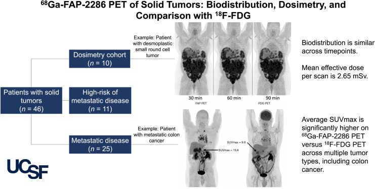
Fibroblast activation protein (FAP), expressed in the tumor microenvironment of a variety of cancers, has become a target of novel PET tracers. The purpose of this report is to evaluate the imaging characteristics of 68Ga-FAP-2286, present the first-to our knowledge-dosimetry analysis to date, and compare the agent with 18F-FDG and FAPI compounds. Methods: Patients were administered 219 ± 43 MBq of 68Ga-FAP-2286 and scanned after 60 min. Uptake was measured in up to 5 lesions per patient and within the kidneys, spleen, liver, and mediastinum (blood pool). Absorbed doses were evaluated using MIM Encore and OLINDA/EXM version 1.1 using the International Commission on Radiological Protection publication 103 tissue weighting factor. Results: Forty-six patients were imaged with 68Ga-FAP-2286 PET. The highest average uptake was seen in sarcoma, cholangiocarcinoma, and colon cancer. The lowest uptake was found in lung cancer and testicular cancer. The average SUVmax was significantly higher on 68Ga-FAP-2286 PET than on 18F-FDG PET in cholangiocarcinoma (18.2 ± 6.4 vs. 9.1 ± 5.0, P = 0.007), breast cancer (11.1 ± 6.8 vs. 4.1 ± 2.2, P < 0.001), colon cancer (13.8 ± 2.2 vs. 7.6 ± 1.7, P = 0.001), hepatocellular carcinoma (9.3 ± 3.5 vs. 4.7 ± 1.3, P = 0.01), head and neck cancer (11.3 ± 3.5 vs. 7.6 ± 5.5, P = 0.04), and pancreatic adenocarcinoma (7.4 ± 1.8 vs. 3.7 ± 1.0, P = 0.01). The total-body effective dose was estimated at 1.16E-02 mSv/MBq, with the greatest absorbed organ dose in the urinary bladder wall (9.98E-02 mGy/MBq). Conclusion: 68Ga-FAP-2286 biodistribution, dosimetry, and tumor uptake were similar to those of previously reported FAPI compounds. Additionally,68Ga-FAP-2286 PET had consistently higher uptake than 18F-FDG PET. These results are especially promising in the setting of small-volume disease and differentiating tumor from inflammatory uptake.
Keywords: FAP-2286; PET; dosimetry; fibroblast activation protein.
© 2024 by the Society of Nuclear Medicine and Molecular Imaging.
Figures


References
-
- Chen H, Pang Y, Wu J, et al. . Comparison of [68Ga]Ga-DOTA-FAPI-04 and [18F] FDG PET/CT for the diagnosis of primary and metastatic lesions in patients with various types of cancer. Eur J Nucl Med Mol Imaging. 2020;47:1820–1832. - PubMed
-
- Zhao L, Pang Y, Luo Z, et al. . Role of [68Ga]Ga-DOTA-FAPI-04 PET/CT in the evaluation of peritoneal carcinomatosis and comparison with [18F]-FDG PET/CT. Eur J Nucl Med Mol Imaging. 2021;48:1944–1955. - PubMed
Publication types
MeSH terms
Substances
Grants and funding
LinkOut - more resources
Full Text Sources
Medical
Miscellaneous