Review
. 2024 May 2;76(3):358-387.
doi: 10.1124/pharmrev.123.000831.

Adrenoceptor Desensitization: Current Understanding of Mechanisms

Affiliations
Review

Adrenoceptor Desensitization: Current Understanding of Mechanisms

Dina Maaliki et al. Pharmacol Rev. .

Abstract

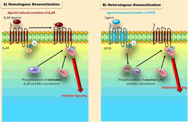
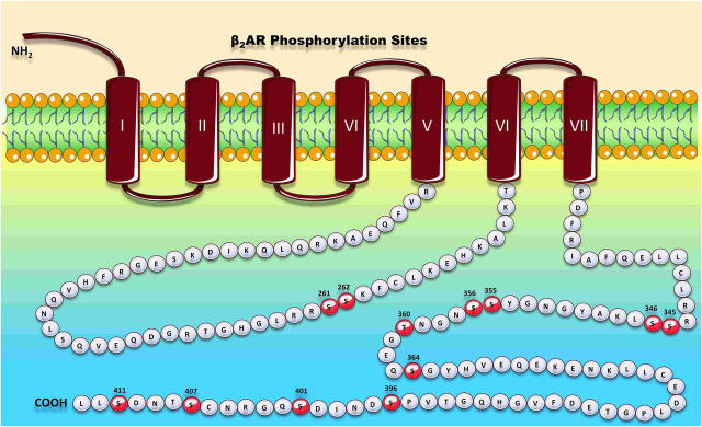
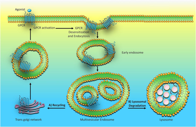
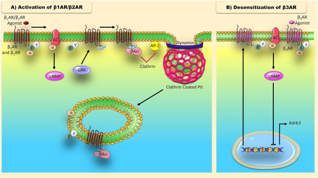
G-protein coupled receptors (GPCRs) transduce a wide range of extracellular signals. They are key players in the majority of biologic functions including vision, olfaction, chemotaxis, and immunity. However, as essential as most of them are to body function and homeostasis, overactivation of GPCRs has been implicated in many pathologic diseases such as cancer, asthma, and heart failure (HF). Therefore, an important feature of G protein signaling systems is the ability to control GPCR responsiveness, and one key process to control overstimulation involves initiating receptor desensitization. A number of steps are appreciated in the desensitization process, including cell surface receptor phosphorylation, internalization, and downregulation. Rapid or short-term desensitization occurs within minutes and involves receptor phosphorylation via the action of intracellular protein kinases, the binding of β-arrestins, and the consequent uncoupling of GPCRs from their cognate heterotrimeric G proteins. On the other hand, long-term desensitization occurs over hours to days and involves receptor downregulation or a decrease in cell surface receptor protein level. Of the proteins involved in this biologic phenomenon, β-arrestins play a particularly significant role in both short- and long-term desensitization mechanisms. In addition, β-arrestins are involved in the phenomenon of biased agonism, where the biased ligand preferentially activates one of several downstream signaling pathways, leading to altered cellular responses. In this context, this review discusses the different patterns of desensitization of the α 1-, α 2- and the β adrenoceptors and highlights the role of β-arrestins in regulating physiologic responsiveness through desensitization and biased agonism. SIGNIFICANCE STATEMENT: A sophisticated network of proteins orchestrates the molecular regulation of GPCR activity. Adrenoceptors are GPCRs that play vast roles in many physiological processes. Without tightly controlled desensitization of these receptors, homeostatic imbalance may ensue, thus precipitating various diseases. Here, we critically appraise the mechanisms implicated in adrenoceptor desensitization. A better understanding of these mechanisms helps identify new druggable targets within the GPCR desensitization machinery and opens exciting therapeutic fronts in the treatment of several pathologies.


Figures

Fig. 1
Fig. 1
Homologous and heterologous desensitization of GPCRs. The desensitization of GPCRs can be classified as homologous or heterologous. (A) Homologous desensitization of a GPCR is ligand dependent. In this type of desensitization, the receptor is phosphorylated by GRKs, which preferentially modify active agonist-occupied receptors. Phosphorylation promotes β-Arr binding, which prevents G protein recruitment and attenuates signaling. (B) Heterologous desensitization is ligand independent. This type of desensitization is mediated by second messenger-dependent protein kinases (PKA and PKC). Within a cell, nearby receptors that lead to activation of second messengers like cAMP can stimulate PKA and PKC. PKA and PKC then phosphorylate any agonist-occupied and unoccupied receptors in their vicinity that contain the consensus phosphorylation sites for these protein kinases.
Fig. 2
Fig. 2
Localization of the β2-AR phosphorylation sites.
Fig. 3
Fig. 3
GPCR sorting itineraries. After receptor internalization, clathrin coated vesicles (CCVs) containing the GPCRs fuse with early endosomes, which then mature into late and MVEs. The GPCR is then sorted into alternative trafficking pathways: (A) It can be dephosphorylated and recycled to the plasma membrane where it can reinitiate signaling (resensitization), or (B) it can be sorted to lysosomes where it is rapidly degraded (downregulation).
Fig. 4
Fig. 4
Illustration of the βAR signaling cycle- (A) The β1- and β2-ARs: Agonist-induced stimulation of the β-ARs leads to the recruitment and activation of heterotrimeric G proteins. Activated G proteins dissociate and the Gαs subunit stimulates adenylyl cyclase-dependent generation of cAMP, while the Gβγ subunits promote other signaling cascades. Protein kinases such as GRKs phosphorylate-activated β-ARs, facilitating their engagement with β-Arr. Active β-Arrs then interact with clathrin and AP2, which are key constituents of the endocytic machinery, driving receptor uptake into clathrin-coated pits. Clathrin-coated pits pinch off into clathrin coated vesicles (CCVs) through the action of dynamin. (B) β3-AR: Agonist-induced activation of the β3-AR stimulates Gs activation and subsequent cAMP generation. Increased cAMP mediates β3-AR desensitization through suppression of receptor mRNA levels, which reduces receptor surface density.
Fig. 5
Fig. 5
Illustration of the β1-AR indicating the observed phosphorylation sites.
Fig. 6
Fig. 6
Figure illustrating the key phosphorylation sites for the α1A-AR.
Fig. 7
Fig. 7
Figure illustrating the key phosphorylation sites for the α1B-AR.
Fig. 8
Fig. 8
Localization of the α1D-AR phosphorylation sites.
Fig. 9
Fig. 9
Figure Illustrating key phosphorylation sites for the α2A-AR. The sequence LEESSSS-DHAERPPG present in the third intracellular loop is a substrate for GRK-mediated phosphorylation.
Fig. 10
Fig. 10
Localization of the α2B-AR phosphorylation sites. Deletion of three glutamic acid residues in the intracellular tail decreases desensitization of the receptor.
Fig. 11
Fig. 11
β-Arr- and/or G protein-biased agonists versus balanced agonists.
Fig. 12
Fig. 12
Classic versus endosomal GPCR signaling. (A) Classic view of GPCR activation (classic signaling): Interaction of a GPCR with an agonist initiates signaling through the recruitment heterotrimeric G proteins. The activated Gα subunit stimulates downstream signaling mediators, which results in transient signaling at the plasma membrane. This signaling is attenuated upon GPCR phosphorylation by protein kinases such as GRK and subsequent β-Arr binding. Engagement of the GPCR with β-Arr inhibits further G protein recruitment and initiates receptor endocytosis and desensitization. (B) Updated model of GPCR activation (endosomal signaling): Interaction of a GPCR with a ligand can lead to signaling through G proteins or β-Arrs, as well as desensitization and endocytosis through β-Arrs. (1) Different ligands can induce distinct GPCR phosphorylation patterns, which dictate subsequent receptor-β-Arr conformations and functional outcomes. (2) Types of receptor β-Arr conformations include the “tail” and the “core” conformation. In the “tail” conformation, the binding of β-Arr to the phosphorylated GPCR tail leaves the GPCR intracellular core exposed, which allows for sustained G protein interaction. In the “core” conformation, the binding of the β-Arr to the GPCR intracellular core physically hinders further G protein coupling. (3) The “tail” conformation promotes the formation of a super-complex that consists of a single GPCR, β-Arr, and G proteins that can signal from endosomes while undergoing internalization mediated by β-Arr. On the other hand, the “core” conformation terminates G protein signaling and mediates endosomal signaling through β-Arrs.

Similar articles

Cited by

References

    1. Aggarwal A, Esler MD, Socratous F, Kaye DM. Evidence for functional presynaptic alpha-2 adrenoceptors and their down-regulation in human heart failure. J Am Coll Cardiol. 2001;37:1246–1251. - PubMed
    1. Ahn S, Shenoy SK, Wei H, Lefkowitz RJ. Differential kinetic and spatial patterns of β-arrestin and G protein-mediated ERK activation by the angiotensin II receptor. J Biol Chem. 2004;279:35518–35525. - PubMed
    1. Akinaga J, García-Sáinz JA, S Pupo A. Updates in the function and regulation of α1 -adrenoceptors. Br J Pharmacol. 2019;176:2343–2357. - PMC - PubMed
    1. Akinaga J, Lima V, Kiguti LR, Hebeler-Barbosa F, Alcántara-Hernández R, García-Sáinz JA, Pupo AS. Differential phosphorylation, desensitization, and internalization of α1A-adrenoceptors activated by norepinephrine and oxymetazoline. Mol Pharmacol. 2013;83:870–881. - PubMed
    1. Albarrán-Juárez J, Gilsbach R, Piekorz RP, Pexa K, Beetz N, Schneider J, Nürnberg B, Birnbaumer L, Hein L. Modulation of alpha2-adrenoceptor functions by heterotrimeric Galphai protein isoforms. J Pharmacol Exp Ther. 2009;331:35–44. - PMC - PubMed

Publication types

MeSH terms

LinkOut - more resources