. 2024 May 2;15(1):3711.
doi: 10.1038/s41467-024-47948-4.

The complete assembly of human LAT1-4F2hc complex provides insights into its regulation, function and localisation

Affiliations

The complete assembly of human LAT1-4F2hc complex provides insights into its regulation, function and localisation

Di Wu et al. Nat Commun. .

Abstract

The LAT1-4F2hc complex (SLC7A5-SLC3A2) facilitates uptake of essential amino acids, hormones and drugs. Its dysfunction is associated with many cancers and immune/neurological disorders. Here, we apply native mass spectrometry (MS)-based approaches to provide evidence of super-dimer formation (LAT1-4F2hc)2. When combined with lipidomics, and site-directed mutagenesis, we discover four endogenous phosphatidylethanolamine (PE) molecules at the interface and C-terminus of both LAT1 subunits. We find that interfacial PE binding is regulated by 4F2hc-R183 and is critical for regulation of palmitoylation on neighbouring LAT1-C187. Combining native MS with mass photometry (MP), we reveal that super-dimerization is sensitive to pH, and modulated by complex N-glycans on the 4F2hc subunit. We further validate the dynamic assemblies of LAT1-4F2hc on plasma membrane and in the lysosome. Together our results link PTM and lipid binding with regulation and localisation of the LAT1-4F2hc super-dimer.


Conflict of interest statement

C.V.R. is a cofounder of and consultant at OMass Therapeutics. The remaining authors declare no competing interests.

Figures
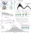
Fig. 1
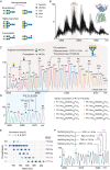
Fig. 1. MS analysis of the LAT1-4F2hc complex.
A Structure of LAT1-4F2hc as reported previously (PDB: 6IRT). Four N-glycans at Asn365, Asn 381, Asn424 and Asn506 are highlighted in purple. The covalently linked 4F2hc-Cys164 (blue) and LAT1-Cys210 (green) are shown. Two lipid-like molecules are shown (pink space filled). B Annotation of potential PTM sites, namely phosphorylation, N-glycosylation and disulfide bonds on 4F2hc and LAT1. C Mass spectrum of the LAT1-4F2hc complex from OGNG micelles. The molecular masses of heterodimeric and an unexpected super-dimeric form of the LAT1-4F2hc complexes are calculated as ~140 kDa and ~280 kDa, respectively. D An expansion of the +26 charge state from the native mass spectrum of the heterodimeric LAT1-4F2hc complex. The Δm/z between the adjacent peaks is calculated and plotted as a scatter plot. The averaged Δm/z (2.81) is plotted as a purple line with the shaded area corresponding to one standard deviation (±0.19, light purple). Each fine structure peak differs by ~73 Da. E Illustration of anticipated N-glycan modifications on LAT1-4F2hc complex. N-glycan branching, extension, sialylation and fucosylation are common features found in the glycome of LAT1-4F2hc. F An illustration of how the combinatorial effects of N-glycan branching, extension, sialylation and fucosylation impact the molecular weight (MW) of the intact glycoprotein. Different combinations of these monosaccharide residues result in repeating 72.95 ± 0.69 Da increments on the intact glycoprotein masses (Supplementary Fig. 2). G Zero-charged spectrum of LAT1-4F2hc. The base peak labelled with a red asterisk is assigned as LAT1-4F2hc with one tri-antennary and three tetra-antennary sialylated N-glycans with four or six additional fucose residues. The N-glycan compositions of other peaks can be inferred based on the mass differences to the base peak.
Fig. 2
Fig. 2. MS analysis of the desialylated LAT1-4F2hc complex.
A Schematic illustration of neuraminidase treatment of N-glycans in which all sialic acid residues are removed from N-glycans. B Native mass spectrum of desialylated LAT1-4F2hc. C Annotation of the N-glycan heterogeneity on desialylated LAT1-4F2hc (charge +27). The major peaks, differing by one GlcNAc1Gal1 unit (365.33 Da), are labelled with P1 – P10. The peaks carrying zero to four antennary fucose residues are annotated with aF0 to aF4, respectively. The P5 proteoform (measured mass of 137027 ± 2 Da) was assigned to LAT1-4F2hc complex with four tetra-antennary core-fucosylated N-glycans (theoretical mass of 137028 Da). D Annotation of antennary fucosylation status of P3 proteoforms. The P3 aF3 and aF4 proteoforms are 74.0 ± 0.7 Da and 214.7 ± 2.2 Da larger than the P4 aF0 proteoform, respectively. E Lipidomics analysis of co-purified phospholipids with LAT1-4F2hc. The identified phospholipids, namely PA, PE, PC, PG, PS and PI are plotted according to their relative abundances (light to dark blue dots). F Annotation of the peaks of the co-purified lipids (PE, PS and PI) with mass shifts (650–900 Da) and their potential overlap with glycoforms of the P4 proteoform.
Fig. 3
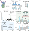
Fig. 3. Native MS analysis reveals endogenous PE binding to the dissociated LAT1-4F2hc complex.
A TCEP treatment reduces the disulfide bond between LAT1 and 4F2hc subunits. B Mass photometry measurements of LAT1-4F2hc assemblies in 36 µM GDN without (control) and with TCEP treatment. The molecular weights of LAT1-4F2hc heterodimer and super-dimer in GDN proteomicelles are ~177 kDa and 341 kDa, respectively. The bar graph shows ratios of LAT1-4F2hc heterodimer and super-dimer without (control) and with TCEP treatment. Bars show mean ± standard deviation from three independent experiments (dots). Source data are provided as a Source Data file. C Native MS analysis of the TCEP-treated LAT1-4F2hc and gas-phase dissociation of the two subunits. The native MS parameters, including capillary temperature (Capillary Temp.), source fragmentation energy (Source Frag.), In-source trapping energy (IST) and HCD energy (HCD) are labelled. D The spectrum of dissociated LAT1 (charge state + 15) reveals one phosphorylation (80.2 ± 1.3 Da) and two lipid adducts (730.1 ± 2.8 Da and 732.5 ± 0.5 Da). E The spectra of dissociated 4F2hc subunit (charge state + 15, top panel) and desialylated LAT1-4F2hc complex (charge state + 27, bottom panel). The corresponding proteoforms of the 4F2hc subunit (P1′ – P9′) and the LAT1-4F2hc heterodimer (P1 – P9) are aligned. The absence of the LAT1 subunit and its associated endogenous lipids results in the decreased abundances of P5 to P9 peaks of the dissociated 4F2hc subunit (the upper spectrum highlighted in green).
Fig. 4
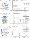
Fig. 4. Mutation of LAT1-4F2hc complexes to investigate lipid binding, the effects of glycosylation and the influence of the LAT1 C-terminus.
A The phospholipid binding site is at the C-terminus of the LAT1 subunit (light green) and the transmembrane segment of 4F2hc (blue). The head group of interfacial phospholipid interacts with 4F2hc-R183. B Native mass spectra of LAT1-4F2hc with the 4F2hc-R183L mutation. No lipid adduct is observed. The palmitoylated proteoform is labelled with a purple circle. C Structural illustration of non-glycosylated LAT1-4F2hc 4 M mutant with N365D/N381D/N424D/N506D. D Native mass spectra of non-glycosylated LAT1-4F2hc 4 M mutant in 20 µM LMNG. Two endogenous lipid adducts (731.3 ± 2.7 Da and 731.8 ± 4.2 Da) are observed. E Structural illustration of LAT1-4F2hc 4M-ΔC mutant with deletion of LAT1 C-terminal helix (highlighted orange). F Native mass spectra of LAT1-4F2hc 4M-ΔC mutant in LMNG. The absence of lipid adducts means that no endogenous lipids are retained. LMNG adduct peaks are highlighted with asterisks.
Fig. 5
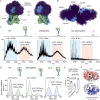
Fig. 5. Native MS and mass photometry analysis of LAT1-4F2hc super-dimerization.
A Simulation of possible N-glycan conformers on LAT1-4F2hc. N-glycan conformers are highlighted in dark purple. B Structural illustration of the modelled structure of LAT1-4F2hc super-dimer with possible N-glycan conformers. The 4F2hc-Asn365 and Asn424 are proximal to the super-dimer interface. The N-glycans fill the interface and mediate hydrophilic interactions between two 4F2hc subunits in the LAT1-4F2hc super-dimer. C Native mass spectra of LAT1-4F2hc WT, desialylated WT and non-glycosylated M4 mutant. The LAT1-4F2hc heterodimer and its super-dimer peaks are highlighted (blue and red, respectively). D Mass photometry analysis of LAT1-4F2hc fully sialylated WT (control), desialylated WT and sialylated WT at pH 5.0. E Bargraphs show the super-dimer/heterodimer ratios of fully sialylated WT (control), desialylated WT and sialylated WT at pH 5.0. Bars show mean ± standard deviation from three independent experiments (dots). Two tailed Student’s t-tests are performed for statistical analysis (p < 0.01 was labelled with two asterisks). Source data are provided as a Source Data file. F Surface electrostatic potential of the super-dimer interface of 4F2hc subunit at pH 7.0 and pH 5.0.
Fig. 6
Fig. 6. Probing the endogenous LAT1-4F2hc assemblies.
A Western blotting analysis of the in vivo crosslinked (XL) endogenous LAT1-4F2hc in HeLa cells. An anti-LAT1 antibody was used to probe the endogenous LAT1-4F2hc assemblies without BS3 (-) and with BS3 (+) treatments. The experiment was repeated once. Source data are provided as a Source Data file. B Correlation of the mRNA expression levels of 4F2hc (SLC3A2) and its associated members of the SLC7A family (SLC7A5, SLC7A6, SLC7A7, SLC7A8, SLC7A9, SLC7A10) from human tissue and cell lines. The mRNA expression levels (measured in normalized transcripts per million, nTPM) of 4F2hc and SLC7As from 50 human tissues and 1205 cell lines were plotted as a scatter plot. The linear-fit trendline is plotted as a dark blue line. The shaded areas corresponding to one and two standard deviations (σ) of the linear regression are highlighted (dark blue and light blue, respectively). Source data are provided as a Source Data file. C Expression levels of SLC3A2 and SLC7A5/6/7/8/10/11 in HeLa cell lines. Source data are provided as a Source Data file. D Western blotting of the endogenous LAT1-4F2hc assemblies in HeLa cells without and with dithiothreitol (DTT) treatment. The experiments were repeated once. Source data are provided as a Source Data file. E Model of the dynamic assemblies of LAT1-4F2hc super-dimer with highlighted key findings. The LAT1 homo-dimer and LAT1-4F2hc super-dimer structures are proposed based on AlphaFold-Multimer and homology-modelling.

Similar articles

Cited by

References

    1. Scalise, M., Galluccio, M., Console, L., Pochini, L. & Indiveri, C. The human SLC7A5 (LAT1): The intriguing histidine/large neutral amino acid transporter and its relevance to human health. Front. Chem.6, 1–12 (2018). 10.3389/fchem.2018.00243 - DOI - PMC - PubMed
    1. Wagner, C. A., Lang, F. & Bröer, S. Function and structure of heterodimeric amino acid transporters. Am. J. Physiol. Cell Physiol.281, C1077–C1093 (2001). 10.1152/ajpcell.2001.281.4.C1077 - DOI - PubMed
    1. Napolitano, L. et al. LAT1 is the transport competent unit of the LAT1/CD98 heterodimeric amino acid transporter. Int. J. Biochem. Cell Biol.67, 25–33 (2015). 10.1016/j.biocel.2015.08.004 - DOI - PubMed
    1. Campbell, W. A. & Thompson, N. L. Overexpression of LAT1/CD98 light chain is sufficient to increase system L-amino acid transport activity in mouse hepatocytes but not fibroblasts. J. Biol. Chem.276, 16877–16884 (2001). 10.1074/jbc.M008248200 - DOI - PubMed
    1. Boado, R. J. et al. Site-directed mutagenesis of cysteine residues of large neutral amino acid transporter LAT1. Biochim. Biophys. Acta1715, 104–110 (2005). 10.1016/j.bbamem.2005.07.007 - DOI - PubMed

Publication types

MeSH terms

Substances