. 2024 May 2;14(1):10054.
doi: 10.1038/s41598-024-60459-y.

Cognitive impairment and hippocampal neuronal damage in β-thalassaemia mice

Affiliations

Cognitive impairment and hippocampal neuronal damage in β-thalassaemia mice

Nuttanan Pholngam et al. Sci Rep. .

Abstract

β-Thalassaemia is one of the most common genetic diseases worldwide. During the past few decades, life expectancy of patients has increased significantly owing to advance in medical treatments. Cognitive impairment, once has been neglected, has gradually become more documented. Cognitive impairment in β-thalassaemia patients is associated with natural history of the disease and socioeconomic factors. Herein, to determined effect of β-thalassaemia intrinsic factors, 22-month-old β-thalassaemia mouse was used as a model to assess cognitive impairment and to investigate any aberrant brain pathology in β-thalassaemia. Open field test showed that β-thalassaemia mice had decreased motor function. However, no difference of neuronal degeneration in primary motor cortex, layer 2/3 area was found. Interestingly, impaired learning and memory function accessed by a Morris water maze test was observed and correlated with a reduced number of living pyramidal neurons in hippocampus at the CA3 region in β-thalassaemia mice. Cognitive impairment in β-thalassaemia mice was significantly correlated with several intrinsic β-thalassaemic factors including iron overload, anaemia, damaged red blood cells (RBCs), phosphatidylserine (PS)-exposed RBC large extracellular vesicles (EVs) and PS-exposed medium EVs. This highlights the importance of blood transfusion and iron chelation in β-thalassaemia patients. In addition, to improve patients' quality of life, assessment of cognitive functions should become part of routine follow-up.

Keywords: Aged mouse model; Behavior test; Brain pathology; Cognitive impairment; Hippocampus; β-thalassaemia.


Conflict of interest statement

The authors declare no competing interests.

Figures

Figure 1
Figure 1
The β-thalassaemia mice had cognitive impairment. Two cognitive functions including motor activity and spatial learning and memory of 22-month-old wild type (WT) and β-thalassaemia (BKO) mice were investigated by (AC) open field tests and (DG) Morris water maze (MWM) tests, respectively. (A) Mouse trajectory path movement in the open field test. The open field test showed decreased motor activity in β-thalassaemia mice indicated by (B) total distance and (C) mean speed. Impaired spatial learning and memory of β-thalassaemia mice was elucidated by MWM tests. (D) Mouse swimming tracks in the MWM tests. The learning curve of mice during the learning phase in the MWM was assessed for (E) latency to find the target platform and (F) distance to the target platform. (G) Percentages of time spent in the target quadrant at probe trial test. Data presents as mean ± SEM. *Statistically significant difference between groups at P < 0.05. ns not significant difference between groups.
Figure 2
Figure 2
No neuronal degeneration in primary motor cortex, layer 2/3 of β-thalassaemia mice. Brains from (A) 22-month-old wild type (WT) mice and (B) 22-month-old β-thalassaemia (BKO) mice were stained with haematoxylin and eosin (H&E) for anatomical analysis (left panel), Nissl stain for counting living and dark neuron cells (middle panel) and Perls’ Prussian blue stain for the evaluation of iron deposition (right panel). Scale bar is 200 μm. (C) Total neuron cells, (D) living neurons and (E) dark neuron in primary motor cortex, layer 2/3 from left- and right-side of brain were counted and identified using Nissl stain (Supplementary Figs. S3, S4). Data presents as mean ± SEM.
Figure 3
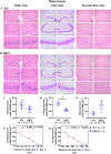
Figure 3
Decreased living pyramidal cells in hippocampal CA3 region of β-thalassaemia mice. Histopathological analysis of the hippocampus from (A) 22-month-old wild type (WT) mice and (B) 22-month-old β-thalassaemia (BKO) mice stained with haematoxylin and eosin (H&E) for anatomical analysis (left panel), Nissl staining for counting living and dark neurons (middle panel) and Perls’ Prussian blue staining for the evaluation of iron deposition (right panel). Scale bar is 200 μm. The β-thalassaemia mice have decreased living pyramidal cells in hippocampal CA3 region. (C) Total pyramidal neurons, (D) living neurons and (E) dark neurons in the hippocampal CA3 region from left- and right-side of the brain were counted and identified as living and dark neurons using Nissl stain (Supplementary Figs. S5–S7). The living pyramidal cells in the hippocampal CA3 region is correlated with cognitive function. Spearman’s rho correlation coefficient analysis between living pyramidal cells in the hippocampal CA3 region and (F) difference time of latency to target between day 5 and day 2 and (G) difference of distance to target between day 5 and day 2. Data presented as mean ± SEM. *Statistically significant difference between groups at P < 0.05.
Figure 4
Figure 4
Clinical pathology of β-thalassaemia mice. β-Thalassaemia (BKO) and wild type (WT) mice were evaluated for (A) liver tissue iron and anaemia markers including (B) red blood cell (RBC) count, (C) haemoglobin (Hb) and (D) haematocrit (Hct); and damaged RBC index including (E) phosphatidylserine (PS)-exposed RBCs, (F) PS-exposed RBC large extracellular vesicles (EVs) and (G) PS-exposed medium EVs. Data presents as mean ± SD. *Statistically significant difference between groups at P < 0.05.
Figure 5
Figure 5
Cognitive impartment correlated with iron overload, anaemia and RBC damage in β-thalassaemia mice. Spearman’s rho correlation coefficient was used to analysed the relationship between liver tissue iron and behavior in spatial learning and memory tests using the MWM test including (A) different time of latency to target between day 5 and day 2, (B) different distance to target between day 5 and day 2, and (C) percentages of time in target quadrant. Percentages of time in target quadrant were correlated with (D) RBC count, (E) haemoglobin (Hb), (F) haematocrit (Hct), (G) phosphatidylserine (PS)-exposed RBCs, (H) PS-exposed RBC large extracellular vesicles (EVs) and (I) PS-exposed medium EVs.

References

    1. Taher AT, Musallam KM, Cappellini M. D. β-Thalassemias. N. Engl. J. Med. 2021;384:727–743. doi: 10.1056/NEJMra2021838. - DOI - PubMed
    1. Haghpanah S, Karimi M. Cerebral thrombosis in patients with β-thalassemia: A systematic review. Blood Coagul. 2012;23:212–217. doi: 10.1097/MBC.0b013e3283502975. - DOI - PubMed
    1. Karimi M, Bagheri H, Rastgu F, Rachmilewitz EA. Magnetic resonance imaging to determine the incidence of brain ischaemia in patients with β-thalassaemia intermedia. Thromb. Haemost. 2010;103:989–993. doi: 10.1160/TH09-09-0661. - DOI - PubMed
    1. Pazgal I, Inbar E, Cohen M, Shpilberg O, Stark P. High incidence of silent cerebral infarcts in adult patients with β-thalassemia major. Thromb. Res. 2016;144:119–122. doi: 10.1016/j.thromres.2016.06.010. - DOI - PubMed
    1. Taher AT, et al. Asymptomatic brain magnetic resonance imaging abnormalities in splenectomized adults with thalassemia intermedia. J. Thromb. Haemost. 2010;8:54–59. doi: 10.1111/j.1538-7836.2009.03651.x. - DOI - PubMed

Publication types

MeSH terms