. 2024 May 3;43(1):134.
doi: 10.1186/s13046-024-03046-3.

CXCR6-positive circulating mucosal-associated invariant T cells can identify patients with non-small cell lung cancer responding to anti-PD-1 immunotherapy

Affiliations

CXCR6-positive circulating mucosal-associated invariant T cells can identify patients with non-small cell lung cancer responding to anti-PD-1 immunotherapy

Jingjing Qu et al. J Exp Clin Cancer Res. .

Abstract

Background: Mucosal-associated invariant T (MAIT) cells have been reported to regulate tumor immunity. However, the immune characteristics of MAIT cells in non-small cell lung cancer (NSCLC) and their correlation with the treatment efficacy of immune checkpoint inhibitors (ICIs) remain unclear.

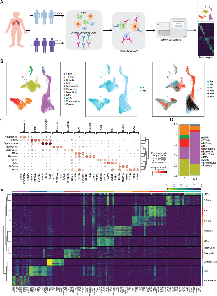
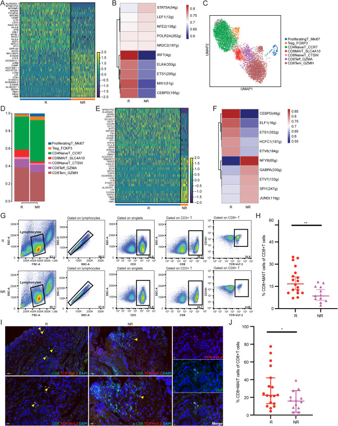
Patients and methods: In this study, we performed single-cell RNA sequencing (scRNA-seq), flow cytometry, and multiplex immunofluorescence assays to determine the proportion and characteristics of CD8+MAIT cells in patients with metastatic NSCLC who did and did not respond to anti-PD-1 therapy. Survival analyses were employed to determine the effects of MAIT proportion and C-X-C chemokine receptor 6 (CXCR6) expression on the prognosis of patients with advanced NSCLC.

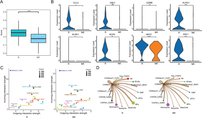
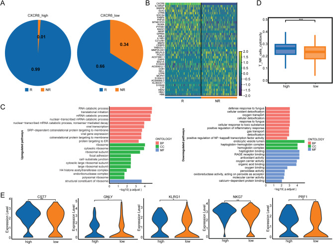
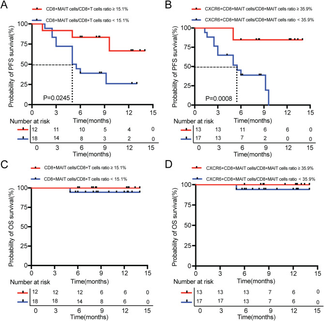
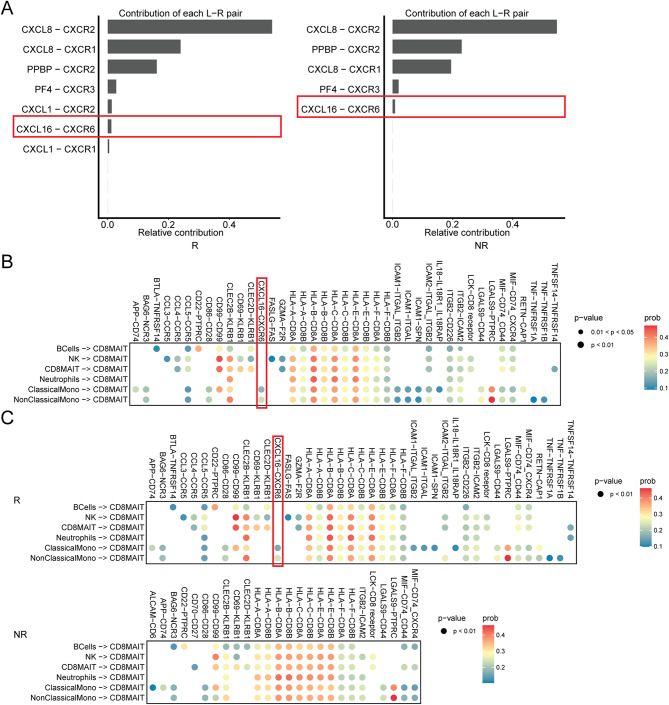
Results: The proportion of activated and proliferating CD8+MAIT cells were significantly higher in responders-derived peripheral blood mononuclear cells (PBMCs) and lung tissues before anti-PD-1 therapy, with enhanced expression of cytotoxicity-related genes including CCL4, KLRG1, PRF1, NCR3, NKG7, GZMB, and KLRK1. The responders' peripheral and tumor-infiltrating CD8+MAIT cells showed an upregulated CXCR6 expression. Similarly, CXCR6+CD8+MAIT cells from responders showed higher expression of cytotoxicity-related genes, such as CST7, GNLY, KLRG1, NKG7, and PRF1. Patients with ≥15.1% CD8+MAIT cells to CD8+T cells ratio and ≥35.9% CXCR6+CD8+MAIT cells to CD8+MAIT cells ratio in peripheral blood showed better progression-free survival (PFS) after immunotherapy. The role of CD8+MAIT cells in lung cancer immunotherapy was potentially mediated by classical/non-classical monocytes through the CXCL16-CXCR6 axis.

Conclusion: CD8+MAIT cells are a potential predictive biomarker for patients with NSCLC responding to anti-PD-1 therapy. The correlation between CD8+MAIT cells and immunotherapy sensitivity may be ascribed to high CXCR6 expression.

Keywords: CXCR6; Circulating mucosal-associated invariant T cells; Immunotherapy; Non-small cell lung cancer; Single-cell RNA-sequencing.


Conflict of interest statement

The authors declare that there are no conflicts of interest.

Figures

Fig. 1
Fig. 1
A higher proportion of T cells in responders. (A) The flowchart of scRNA-seq (peripheral blood samples of NSCLC patients were collected from 3 responders and 3 non-responders at baseline). (B) UMAP analysis plot showing cell clusters (left), classification according to anti-PD-1 therapy response (middle) and the derivation of samples (right). (C) The expression of canonical marker genes in different cell subtypes. (D) The proportion of cell subtypes in responders and non-responders. (E) Differentially expressed genes of different cell subtypes R: Responders; NR: Non-Responders. GMPs: Granulocyte-monocyte progenitor cells; NK: Natural killer cells; MPs: Mononuclear phagocytes; pDCs: Plasmacytoid dendritic cells
Fig. 2
Fig. 2
CD8+MAIT cells showed a significantly increased proportion in responders. (A) Differentially expressed genes between responders and non-responders-derived T cells. (B) Scenic analysis for responders and non-responders-derived T cells. (C) UMAP analysis plot showing T-cell subtypes. (D) The proportion of T-cell subtypes in responders and non-responders. (E) Differentially expressed genes between responders and non-responders in CD8+MAIT cells. (F) Scenic analysis for responders and non-responders-derived CD8+MAIT cells. (G) Flow cytometric analysis of responders and non-responders-derived PBMCs by labeling CD3, CD8, CD161, TCR α7.2(18 responders vs. 12 non-responders). (H) The percentage of CD8+MAIT cells in responders and non-responders-derived CD8+T cells from cytometric analysis (18.7% VS 10.0%, P = 0.0057). (I) Tumor tissues stained by immunofluorescence for CD8, TCR Vα7.2 in responders and non-responders. (J) Percentage of CD8+MAIT cells in CD8+T cells in responders and non-responders (30.1% VS 15.7%, P = 0.0347)
Fig. 3
Fig. 3
Responders in CD8+MAIT cells had higher expression of cytotoxicity-related genes. (A) Cytotoxicity scoring for responders and non-responders-derived CD8+MAIT cells. (B) Differentially expressed cytotoxic genes between responders and non-responders-derived CD8+MAIT cells. (C) The incoming and outgoing cell-cell interaction strength for responders and non-responders-derived cell subtypes. (D) The outgoing interaction pattern for responders and non-responders-derived CD8+MAIT.
Fig. 4
Fig. 4
CD8+MAIT in responders cohort with higher CXCR6 expression. (A) Pseudo-time analysis for CXCR6 expression in different T-cell subtypes. (B) UMAP analysis for CXCR6 expression in responders and non-responders-derived CD8+MAIT cells. (C) Violin plots for CXCR6 expression in responders and non-responders-derived different T-cell subtypes. (D) Flow cytometric analysis of responders and non-responders-derived PBMCs by labeling CD3, CD8, CD161, TCR α7.2 and CD186 (18 responders vs. 12 non-responders). (E) The percentage of CD186+ (CXCR6+) CD8+MAIT cells in CD8+MAIT cells in responders and non-responders from cytometric analysis(P = 0.041). (F-G) Tumor tissues stained by immunofluorescence for CD8, TCR Vα7.2, CXCR6 in responders and non-responders and the summary data (47.44% VS 26.72%, P = 0.0274).
Fig. 5
Fig. 5
CXCR6 in responders was associated with cytotoxicity of CD8+MAIT cells. (A) The components of CXCR6-high and CXCR6-low CD8+MAIT cells. (B) Differentially expressed genes between CXCR6-high and CXCR6-low CD8+MAIT cells. (C) GO analysis for upregulated (left) and downregulated (right) pathways in CXCR6-high CD8+MAIT cells. (D) Cytotoxicity scoring for CXCR6-high and CXCR6-low CD8+MAIT cells. (E) Differentially expressed cytotoxic genes between CXCR6-high and CXCR6-low CD8+MAIT cells (<0.05)
Fig. 6
Fig. 6
Levels of CD8+MAIT and CXCR6+CD8+MAIT cells before anti-PD-1 therapy identify responder patients. (A) CD8+MAIT cells/CD8+T cells ratio ≥15.1% had a better PFS than those patients with CD8+MAIT cells/CD8+T cells ratio <15.1% (NR vs. 5.3 months, P = 0.0245). (B) CXCR6+CD8+MAIT cells/CD8+MAIT cells ratio ≥35.9% had a better PFS than those patients with CXCR6 + CD8+MAIT cells/CD8+MAIT cells ratio <35.9% (NR vs. 5.5 months, P = 0.0008). (C-D) Kaplan–Meier survival analysis of the OS in patients with CD8+MAIT cells/CD8+T cells ratio ≥15.1% and < 15.1% (C), CXCR6 + CD8+MAIT cells/CD8+MAIT cells ratio ≥35.9% and < 35.9% (D)
Fig. 7
Fig. 7
The role of CD8+MAIT cells in lung cancer immunotherapy may be achieved by CXCR6-CXCL16 axis. (A) The contributions of main ligand-receptor pairs in cell-cell interaction of responders and non-responders-derived CD8+MAIT cells. (B) Ligand-receptor pairs participated in cell-cell interaction of CD8+MAIT cells. (C) Ligand-receptor pairs participated in cell-cell interaction of responders and non-responders-derived CD8+MAIT cells

Similar articles

Cited by

References

    1. Sung H, Ferlay J, Siegel RL, Laversanne M, Soerjomataram I, Jemal A, Bray F. Global Cancer statistics 2020: GLOBOCAN estimates of incidence and Mortality Worldwide for 36 cancers in 185 countries. CA Cancer J Clin. 2021;71(3):209–49. doi: 10.3322/caac.21660. - DOI - PubMed
    1. Inamura K. Lung Cancer: understanding its Molecular Pathology and the 2015 WHO classification. Front Oncol. 2017;7:193. doi: 10.3389/fonc.2017.00193. - DOI - PMC - PubMed
    1. Travis WD, Brambilla E, Nicholson AG, Yatabe Y, Austin JHM, Beasley MB, Chirieac LR, Dacic S, Duhig E, Flieder DB, et al. The 2015 World Health Organization Classification of Lung Tumors: impact of genetic, clinical and radiologic advances since the 2004 classification. J Thorac Oncol. 2015;10(9):1243–60. doi: 10.1097/JTO.0000000000000630. - DOI - PubMed
    1. Lahiri A, Maji A, Potdar PD, Singh N, Parikh P, Bisht B, Mukherjee A, Paul MK. Lung cancer immunotherapy: progress, pitfalls, and promises. Mol Cancer. 2023;22(1):40. doi: 10.1186/s12943-023-01740-y. - DOI - PMC - PubMed
    1. Zhang Y, Zhang Z. The history and advances in cancer immunotherapy: understanding the characteristics of tumor-infiltrating immune cells and their therapeutic implications. Cell Mol Immunol. 2020;17(8):807–21. doi: 10.1038/s41423-020-0488-6. - DOI - PMC - PubMed

MeSH terms

Substances