. 2024 May 2;19(1):34.
doi: 10.1186/s13062-024-00468-z.

LncRNA PCED1B-AS1 mediates miR-3681-3p/MAP2K7 axis to promote metastasis, invasion and EMT in gastric cancer

Affiliations

LncRNA PCED1B-AS1 mediates miR-3681-3p/MAP2K7 axis to promote metastasis, invasion and EMT in gastric cancer

Jia Cao et al. Biol Direct. .

Abstract

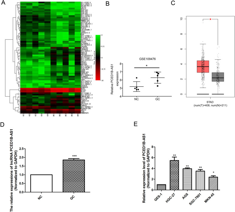
Background: LncRNA PCED1B-AS1 is abnormally expressed in multiple cancers and has been confirmed as an oncogene. Our study aimed to investigate the regulatory mechanism of lncRNA PCED1B-AS1 in gastric cancer.

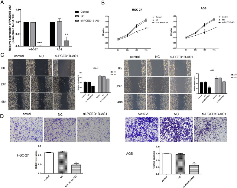
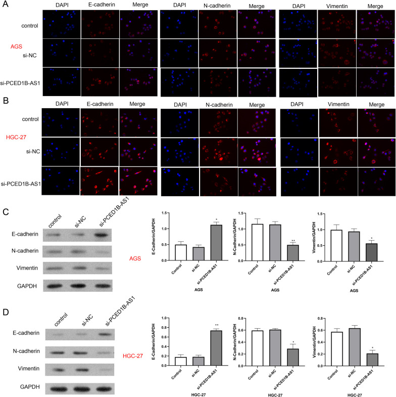
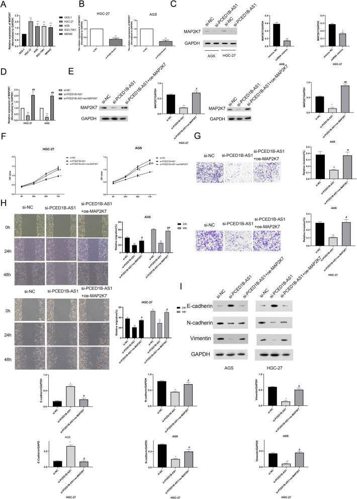
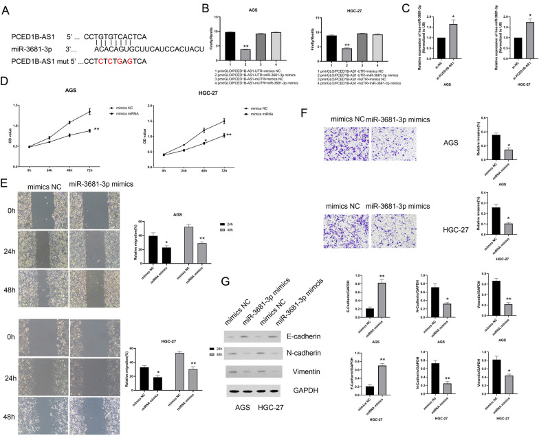
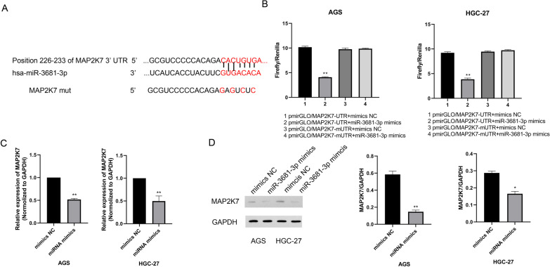
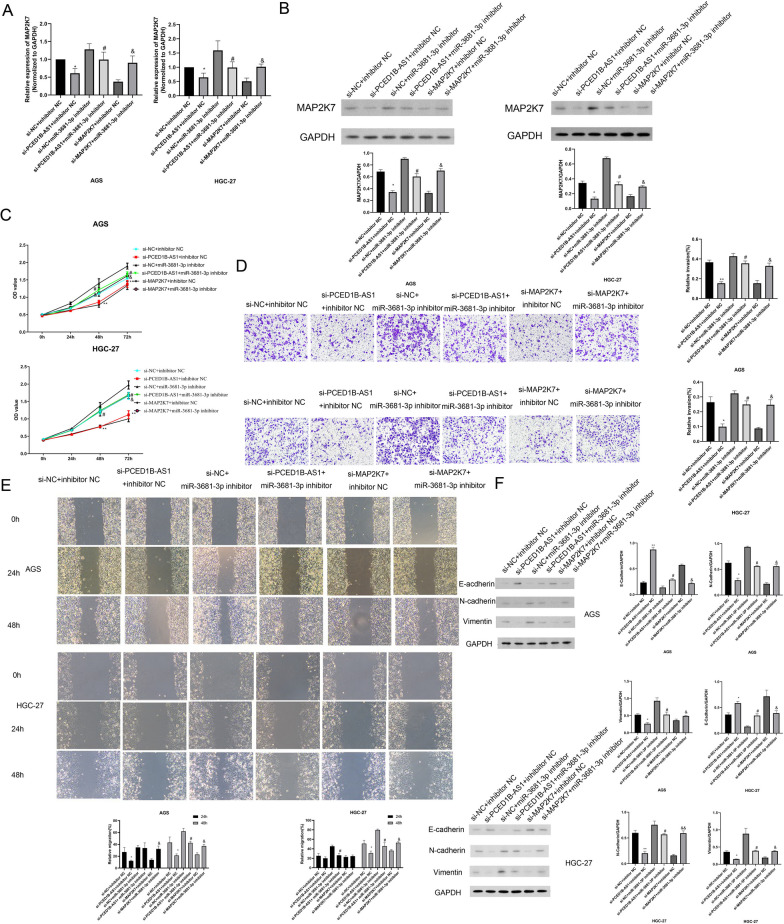
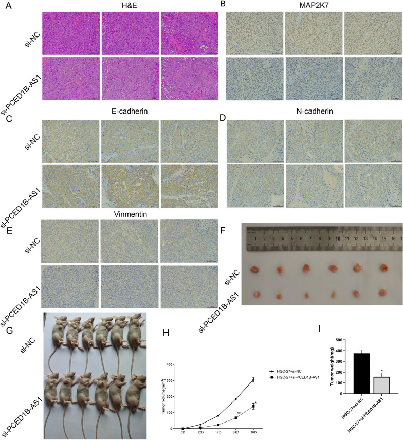
Methods: TCGA database was used to analyze the abnormal expression of lncRNA PCED1B-AS1 in gastric cancer. By database prediction and mass spectrometric analysis, miR-3681-3p and MAP2K7 are potential downstream target molecules of lncRNA PCED1B-AS1 and verified by dual-luciferase report assay. RT-qPCR analysis and western blot were performed to detect the expressions of PCED1B-AS1 and MAP2K7 in gastric cancer cell lines and tissues. CCK-8 kit was applied to measure the cell viability. Wound healing and Transwell experiment were used to detect the migration and invasion. Western blot and immunohistochemical staining were performed to detect the expressions of EMT-related proteins in tissues. The changes of tumor proliferation were detected by xenograft experiment in nude mice.

Results: PCED1B-AS1 expression was higher but miR-3681-3 expression was lower in gastric cancer cell lines or tissues, compared to normal group. Function analysis verified PCED1B-AS1 promoted cell proliferation and inhibited cell apoptosis in gastric cancer cells in vitro and in vivo. LncRNA PCED1B-AS1 could bind directly to miR-3681-3p, and MAP2K7 was found to be a downstream target of miR-3681-3p. MiR-3681-3p mimics or si-MAP2K7 could partly reverse the effect of PCED1B-AS1 on gastric cancer cells.

Conclusion: PCED1B-AS1 accelerated cell proliferation and inhibited cell apoptosis through sponging miR-3681-3p to upregulate MAP2K7 expression in gastric cancer, which indicated PCED1B-AS1/miR-3681-3p/MAP2K7 axis may serve as a potential therapeutic target for gastric cancer.

Keywords: Gastric cancer; MAP2K7; lncRNA PCED1B-AS1; miR-3681-3p.


Conflict of interest statement

All authors declare that there is no any competing interest.

Figures

Fig. 1
Fig. 1
PCED1B-AS1 was overexpressed in gastric cancer tissues. A Heat map showed lncRNA expression levels in tumor tissue and its paired normal tissue. B Genotype-Tissue Expression (GTEx) data predicted the mRNA expression of PCED1B-AS1 in tumor tissue and its paired normal tissue (n = 5). C TCGA database predicted the expression level of PCED1B-AS1 in tumor tissues (n = 408) and normal tissues (n = 211). D RT-qPCR analysis was used to detect the expressions of lncRNA PCED1B-AS1 in the tissues of patients (n = 10). E RT-qPCR analysis was used to detect the levels of PCED1B-AS1 in the gastric cancer cells. The data were presented as mean ± SEM. versus NC/GES-1 group, *P < 0.05, **P < 0.01, ***P < 0.001
Fig. 2
Fig. 2
Knockdown PCED1B-AS1 inhibited cell viability, invasion and migration in gastric cancer cell line. A RT-qPCR analysis was performed to detect the mRNA expression of PCED1B-AS1 in GC cell lines (AGS and HGC-27). B CCK-8 kit was used to assess the effect of PCED1B-AS1 on cell viability in AGS and HGC-27. C Wound healing assay was used to detect the migration in AGS and HGC-27. D Transwell experiment was performed to detect the invasion in AGS and HGC-27. The data were presented as mean ± SEM in three independent experiments. Versus NC group, *P < 0.05, **P < 0.01, n = 3
Fig. 3
Fig. 3
Knockdown PCED1B-AS1 inhibited the EMT process in gastric cancer cell line. Immunofluorescence staining was used to detect the expression of E-cadherin, N-cadherin and Vimentin in AGS cells (A) and HGC cells (B). The nucleus was labeled with DAPI (blue). Western blotting assay was performed to detect the protein expressions of E-cadherin, N-cadherin and Vimentin in AGS cells (C) and HGC-27 cells (D). The data were presented as mean ± SEM in three independent experiments. Versus si-NC group, *P < 0.05, **P < 0.01, n = 3
Fig. 4
Fig. 4
PCED1B-AS1 located in the cytoplasm of gastric cancer cells. A Localization of PCED1B-AS1 in gastric cancer cells was detected by fluorescence in situ hybridization (FISH). Nuclei were stained with DAPI. B RT-qPCR analysis was used to detect the mRNA expression of PCED1B-AS1 in cytoplasm and nucleus. The data were presented as mean ± SEM, n = 3 each group
Fig. 5
Fig. 5
PCED1B-AS1 mediated MAP2K7 to affect the function of gastric cancer cells. A RT-qPCR analysis was performed to detect the mRNA expressions of MAP2K7 in gastric cancer cells. B RT-qPCR analysis was performed to detect the mRNA expressions of MAP2K7 in different groups. C Western blotting assay was used to detect the protein expressions of MAP2K7 in different groups. D After transfection with si-PCED1B-AS1 and oe-MAP2K7, the mRNA expression of MAP2K7 was detected by RT-qPCR analysis. E The protein expressions of MAP2K7 in different groups were detected by western blotting assay. F Cell viability was assessed by CCK-8 kit. G Transwell experiment was used to detect the invasion in different groups. H Wound healing assay was used to detect the migration in different groups. The expressions of E-cadherin, N-cadherin and Vimentin in different groups were detected by western blotting assay. The data were presented as mean ± SEM, versus si-NC group, *P < 0.05, **P < 0.01; versus si-PCED1B-AS1 group, #P < 0.05, ##P < 0.01, n = 3
Fig. 6
Fig. 6
PCED1B-AS1 targeting miR-3681-3p affected gastric cancer cell function. A Predicted binding sites and mutant sites of miR-3681-3p on the PCED1B-AS1 transcript. B Dual luciferase reporter assay demonstrated that both mimics control or miR-3681-3p mimics were separately transfected into AGS and HGC-27 cells with pmirGLO-PCED1B-AS1 (wide-type) or pmirGLO-PCED1B-AS1 (mutant). C RT-qPCR analysis was used to detect the expressions of miR-3681-3p in different groups. D CCK-8 kit was used to detect the cell viability in different groups. E Wound healing assay was performed to detect the migration in different groups. F Transwell experiment was used to detect the invasion in different groups. G The expressions of E-cadherin, N-cadherin and Vimentin in AGS and HGC-27 cells were detected by western blotting assay. The data were presented as mean ± SEM, versus mimics NC group, *P < 0.05, **P < 0.01
Fig. 7
Fig. 7
MAP2K7 acts as a spong of miR-3681-3p in gastric cancer cell. A Predicted binding sites and mutant sites of miR-3681-3p on the MAP2K7 transcript. B Dual luciferase reporter assay demonstrated that both mimics control or miR-3681-3p mimics were separately transfected into AGS and HGC-27 cells with pmirGLO-MAP2K7 (wide-type) or pmirGLO-MAP2K7 (mutant). C RT-qPCR analysis was used to detect the mRNA expressions of MAP2K7 in AGS and HGC-27 cells. D The expressions of MAP2K7 in AGS and HGC-27 cells were detected by western blotting assay. The data were presented as mean ± SEM, versus mimics NC group, *P < 0.05, **P < 0.01, n = 3
Fig. 8
Fig. 8
PCED1B-AS1 mediated miR-3681-3p/MAP2K7 signaling axis to regulate gastric cancer cell function. A RT-qPCR analysis was used to detect the mRNA expressions of MAP2K7 in AGS and HGC-27 cells. B Western blotting assay was performed to detect the expressions of MAP2K7 in AGS and HGC-27 cells. C The cell viability was assessed by CCK-8 kit. D Transwell experiment was performed to detect the invasion in AGS and HGC-27 cells. E Wound healing assay was used to detect the migration in AGS and HGC-27 cells. F The expressions of E-cadherin, N-cadherin and Vimentin in AGS and HGC-27 cells were detected by western blotting assay. The data were presented as mean ± SEM, versus mimics NC group, *P < 0.05, **P < 0.01; versus si-NC + miR-3681-3p inhibitor group, #P < 0.05, ##P < 0.01; versus si-MAP2K7 + inhibitor NC group, &P < 0.05, n = 3
Fig. 9
Fig. 9
Knockdown PCED1B-AS1 inhibited the progression of gastric cancer in mice. A The histopathological changes of gastric cancer were detected by H&E staining. Immunohistochemical staining was used to detect the expression of MAP2K7 (B), E-cadherin (C), N-cadherin (D) and Vimentin (E) in gastric cancer tissues. F Representative tumor tissue. G Representative nude mice are shown. H Statistical analysis of tumor volume (H) and tumor weight (I) from tumors obtained from two groups. The data were presented as mean ± SEM, versus si-NC group, *P < 0.05, **P < 0.01, n = 6

Similar articles

Cited by

References

    1. Joshi S, Badgwell B. Current treatment and recent progress in gastric cancer. CA Cancer J Clin. 2021;71(3):264–279. doi: 10.3322/caac.21657. - DOI - PMC - PubMed
    1. Lazăr D, et al. Prognostic significance of tumor immune microenvironment and immunotherapy: novel insights and future perspectives in gastric cancer. World J Gastroenterol. 2018;24(32):3583–3616. doi: 10.3748/wjg.v24.i32.3583. - DOI - PMC - PubMed
    1. Lin M, et al. Resveratrol-modified mesoporous silica nanoparticle for tumor-targeted therapy of gastric cancer. Bioengineered. 2021;12(1):6343–6353. doi: 10.1080/21655979.2021.1971507. - DOI - PMC - PubMed
    1. Riihimäki M, et al. Metastatic spread in patients with gastric cancer. Oncotarget. 2016;7(32):52307–52316. doi: 10.18632/oncotarget.10740. - DOI - PMC - PubMed
    1. Ahmad I, et al. Long noncoding RNA in myeloid and lymphoid cell differentiation, polarization and function. Cells. 2020;9(2):269. doi: 10.3390/cells9020269. - DOI - PMC - PubMed

Publication types