Review
. 2024 Dec 16;7(12):8040-8058.
doi: 10.1021/acsabm.4c00150. Epub 2024 May 2.

Ultrasound-Controlled Prodrug Activation: Emerging Strategies in Polymer Mechanochemistry and Sonodynamic Therapy

Affiliations
Review

Ultrasound-Controlled Prodrug Activation: Emerging Strategies in Polymer Mechanochemistry and Sonodynamic Therapy

Xuancheng Fu et al. ACS Appl Bio Mater. .

Abstract

Ultrasound has gained prominence in biomedical applications due to its noninvasive nature and ability to penetrate deep tissue with spatial and temporal resolution. The burgeoning field of ultrasound-responsive prodrug systems exploits the mechanical and chemical effects of ultrasonication for the controlled activation of prodrugs. In polymer mechanochemistry, materials scientists exploit the sonomechanical effect of acoustic cavitation to mechanochemically activate force-sensitive prodrugs. On the other hand, researchers in the field of sonodynamic therapy adopt fundamentally distinct methodologies, utilizing the sonochemical effect (e.g., generation of reactive oxygen species) of ultrasound in the presence of sonosensitizers to induce chemical transformations that activate prodrugs. This cross-disciplinary review comprehensively examines these two divergent yet interrelated approaches, both of which originated from acoustic cavitation. It highlights molecular and materials design strategies and potential applications in diverse therapeutic contexts, from chemotherapy to immunotherapy and gene therapy methods, and discusses future directions in this rapidly advancing domain.

Keywords: drug delivery; mechanophores; polymer mechanochemistry; prodrugs; sonochemistry; sonodynamic therapy; sonosensitizers; ultrasound.


Conflict of interest statement

The authors declare no competing financial interest.

Figures

Figure 1
Figure 1
Overview of representative ultrasound–material interaction mechanisms involved in ultrasound-responsive prodrug systems: Section 2 on polymer mechanochemistry strategies, Section 3 on sonodynamic therapy (SDT) strategies, and Section 4 on additional ultrasound-mediated strategies. Texts in gray with dotted outlines represent topics mentioned in this review but not extensively covered.
Figure 2
Figure 2
Ultrasound-induced effects in solutions are primarily from acoustic cavitation: (a) Acoustic field causes pressure variation in the solution and results in acoustic cavitation (the formation, growth, and collapse of bubbles). (b) Rapidly collapsing cavitation bubbles generate rapid liquid flows, transducing mechanical forces to the backbone of polymers near the implosion bubbles. (c) The high-temperature cavitation environment causes small molecules to form radical byproducts, while there is no evidence that the extreme conditions found in cavitation bubbles contribute to polymer degradation in nonaqueous liquids because the polymer chains have negligible vapor pressure and are unlikely to be found at the bubble interface. In aqueous solutions, pyrolysis can occur to hydrophobic polymers concentrate at the bubble–air interface. Reproduced with permission from ref (25). Copyright 2009, American Chemical Society.
Figure 3
Figure 3
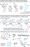
(a) (Sono)mechanochemical activation of furan-maleimide mechanophores triggers the release of payload molecules via a retro-Diels–Alder/fragmentation cascade. (b) Structural modification offers a library of furan-maleimide mechanophores capable of releasing cargo molecules bearing different functional groups. (c) A multimechanophore polymer design incorporating a nonscissile mechanophore enables (sono)mechanically triggered release of hundreds of cargo molecules per chain. Adapted with permission from refs ( and 40). Copyright 2021, American Chemical Society. Copyright 2024, American Chemical Society.
Figure 4
Figure 4
Gas vesicles (GVs)-mediated sonomechanical activation of a CPT-releasing mechanoresponsive PMSEA polymer released the anticancer drug CPT, which exhibited expected cytotoxicity to cancer cells. Sonomechanical activation was performed under physiological conditions using biocompatible focused ultrasound at 330 kHz. Adapted with permission from ref (33). Copyright 2023, The National Academy of Sciences.
Figure 5
Figure 5
(a) Ultrasound-induced scission of disulfide-centered polymers generated thiols that subsequently triggered the release of drug or reporting molecules. Thiol intermediates underwent (A1) intermolecular Michael addition to initiate a retro Diels–Alder reaction and release the furylated doxorubicin payload, (A2) intramolecular 5-exo-trig cyclization to release CPT, and (A3) intramolecular 5-exo-trig cyclization to release CPT and fluorescent umbelliferone simultaneously. (b) Ultrasound mechanochemically induced the cleavage of disulfide cross-linkers in microgels, resulting in a Michael addition to a DA adduct that released a furan dansyl or trimethoprim. Adapted with permission from refs ( and 44). Copyright 2022, Royal Society of Chemistry. Copyright 2022, John Wiley and Sons.
Figure 6
Figure 6
(a) NeoB was loaded into polyaptamers, inhibiting its biological activity. Sonomechanical stretching and scission released NeoB, activating its antibiotic activity. (b) From an enzyme-aptamer complex thrombin@pTBA15, ultrasound triggered the release of thrombin which catalyzed fibrinogen formation from fibrin. (c) Ultrasound treatment induced the disassembly of AuNP/aptamer/thrombin aggregates, leading to the release of active thrombin. Adapted with permission from refs (, , and 49). Copyright 2021, Springer Nature. Copyright 2022, Royal Society of Chemistry. Copyright 2021, John Wiley and Sons.
Figure 7
Figure 7
Sonomechanical force stretched the Au-DNA dimer structures to achieve DOX release. Gold nanoparticles (AuNPs) acted as a transmitter of the sonodynamic shear force, converting double-stranded DNA (the mechanophore) into single stranded. Adapted with permission from ref (50). Copyright 2022, Royal Society of Chemistry.
Figure 8
Figure 8
Structure of the mechanoresponsive cross-linker containing an azo group, and the sonomechanical generation free radicals and ROS from this cross-linked hydrogel. Sonomechanochemically generated ROS leads to different modes of chemical and biological responses. Adapted with permission from ref (35). Copyright 2022, The National Academy of Sciences.
Figure 9
Figure 9
Sound field (top), bubble radius (middle), and light output (bottom) vs time for a sonoluminescencing bubble under 22.3 kHz ultrasound. Sonoluminescence occurred at the most compressed phase of bubble collapsing. Reproduced with permission from ref (81). Copyright 1992, Acoustical Society of America.
Scheme 1
Scheme 1. Representative Structures of Organic Sonosensitizers
Figure 10
Figure 10
(a–c) Structures of semiconducting polymer nanoparticles developed by Pu and co-workers, and schematic illustration of immunomodulator prodrug activation mediated by ultrasound-triggered ROS generation. Adapted with permission from refs (, , and 122). Copyright 2022, Nature Portfolio. Copyright 2023, John Wiley and Sons. Copyright 2022, Nature Portfolio.
Figure 11
Figure 11
(a) The structure of a lipid nanoparticle comprising a ROS-activatable prodrug (LA-GEM), a sonosensitizer (Rhein), and a surfactant (DSPE-PEG2k). (b) The structure of an analogous nanoparticle comprising a ROS-activatable amphiphilic prodrug (PTX-TL-PEG1k-NH2), a sonosensitizer (IR780), and a DSPE-based surfactant. Reproduced with permission from refs ( and 128). Copyright 2023, John Wiley and Sons. Copyright 2023, John Wiley and Sons.
Figure 12
Figure 12
Sonodynamic activation of prodrugs through redox reactions. (a) Schematic illustration of Pt(IV) prodrug Pt1’s sonodynamic activation with a hemoglobin sonosensitizer and the proposed mechanism for the reductive activation (right side). (b) Structure of a Pt(IV) prodrug Cyaninplatin and its sonodynamic activity through two synergistic pathways. (c) DHE’s dual function as a sonosensitizer itself to generate ROS and as an activable prodrug to convert into the cytotoxic ethidium (EB) form under ultrasound irradiation. Adapted with permission from refs (, , and 135). Copyright 2023, American Association for the Advancement of Science. Copyright 2023, John Wiley and Sons. Copyright 2023, John Wiley and Sons.
Figure 13
Figure 13
Ultrasound-controlled site-specific bioorthogonal catalytic azide–alkyne cycloaddition reaction produced a triazole drug in situ and triggered robust sonodynamic therapy. Reproduced with permission from ref (137). Copyright 2023, John Wiley and Sons.
Figure 14
Figure 14
SDT systems involving GSH-activatable sonosensitizers. (a) Structures of 1-Zn-PPA and 1-NLG and their proposed coassembly into 1-NPs for sonophotodynamic immunotherapy. (b) A GSH-activated sonosensitizer prodrug was selectively activated at tumor sites for switch-on SDT activity and fluorescence. Adapted with permission from refs ( and 139). Copyright 2023, John Wiley and Sons. Copyright 2023, John Wiley and Sons.
Figure 15
Figure 15
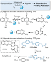
SDT treatments induced hypoxic environments and activated bioreductive prodrugs TPZ (a) and CPT2-Azo (b).
Figure 16
Figure 16
Ultrasound-triggered blue light emission by Lipo@IR780/L012 nanoparticles was harnessed for optogenetic stimulation of opsin-expressing neurons. The mechanism for ultrasound-induced chemiluminescence is illustrated at the bottom. Adapted with permission from ref (52). Copyright 2023, America Chemical Society.
Scheme 2
Scheme 2. (a) Ultrasound-Responsive Prodrug BNN6 Releases Nitric Oxide through Proposed Sonoluminescence-Mediated Photochemical Activation., (b) Urea-Based Prodrug MBU-R Activated by Hydroxy Radicals Generated through Acoustic Cavitation, Leading to Release of Amine-Based Drug Molecule along with Methylene Blue and CO2. (c) IMesNO Prodrug Releases NO via HIFU-Mediated thermolysis (local temperature increase)

Similar articles

Cited by

References

    1. Albert A. Chemical Aspects of Selective Toxicity. Nature 1958, 182 (4633), 421–423. 10.1038/182421a0. - DOI - PubMed
    1. Ding C.; Chen C.; Zeng X.; Chen H.; Zhao Y. Emerging Strategies in Stimuli-Responsive Prodrug Nanosystems for Cancer Therapy. ACS Nano 2022, 16 (9), 13513–13553. 10.1021/acsnano.2c05379. - DOI - PubMed
    1. Bargakshatriya R.; Pramanik S. K. Stimuli-Responsive Prodrug Chemistries for Cancer Therapy. ChemBioChem. 2023, 24 (18), e202300155.10.1002/cbic.202300155. - DOI - PubMed
    1. Maresca D.; Lakshmanan A.; Abedi M.; Bar-Zion A.; Farhadi A.; Lu G. J.; Szablowski J. O.; Wu D.; Yoo S.; Shapiro M. G. Biomolecular Ultrasound and Sonogenetics. Annu. Rev. Chem. Biomol. Eng. 2018, 9 (1), 229–252. 10.1146/annurev-chembioeng-060817-084034. - DOI - PMC - PubMed
    1. Wood A. K. W.; Sehgal C. M. A Review of Low-Intensity Ultrasound for Cancer Therapy. Ultrasound Med. Biol. 2015, 41 (4), 905–928. 10.1016/j.ultrasmedbio.2014.11.019. - DOI - PMC - PubMed

Publication types

LinkOut - more resources