

The Effects of Ultrasonic Scaling and Air-Abrasive Powders on the Topography of Implant Surfaces: Scanning Electron Analysis and In Vitro Study
- PMID: 38698614
- PMCID: PMC11479722
- DOI: 10.1055/s-0044-1782190
The Effects of Ultrasonic Scaling and Air-Abrasive Powders on the Topography of Implant Surfaces: Scanning Electron Analysis and In Vitro Study
Abstract
Objectives: This in vitro study aimed to investigate the impact of bicarbonate air-abrasive powders and ultrasonic scaling with stainless steel tips on the micro- and nanotopography and roughness of three different implant-abutment junction titanium surfaces.
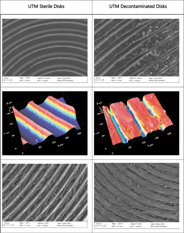
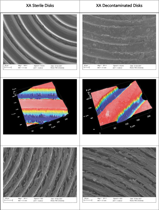
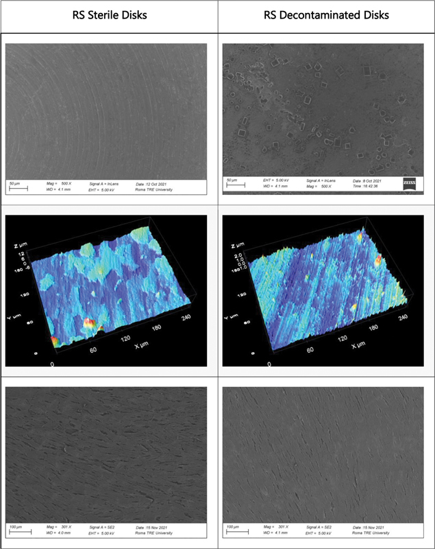
Materials and methods: Three types of sterile and decontaminated titanium surfaces (RS, UTM, XA) were used for analysis. Nine disks per surface type were subjected to micro- and nanotopography analysis, scanning electron microscopy (SEM), roughness analysis, and fibroblast cultivation. Ultrasonic debridement and air polishing were performed on the surfaces. Human dermal fibroblasts were cultured on the surfaces for 5 days.
Statistical analysis: Data analysis adhered to ISO 25178 standards for surface texture assessment. SEM micrographs were used to reconstruct areas for extracting roughness parameters. Excel and Mex 6.0 software were utilized for quantitative and stereoscopic analysis.
Results: The study found varying effects on surface roughness posttreatment. RS Disco samples exhibited higher surface roughness compared with UTM and XA samples, both in average and nanoscale roughness. Decontamination led to increased surface roughness for all samples, particularly RS Disco. Fibroblast growth tests revealed enhanced cell network formation on decontaminated discs, possibly due to increased nanoscale roughness or the presence of bicarbonate salts.
Conclusion: The study underscores the complex interplay between surface topography, microbial biofilm, and treatment efficacy in peri-implant disease management. While smoother surfaces may resist biofilm accumulation, increased nanoscale roughness postdecontamination can enhance fibroblast attachment and soft tissue integration. This dichotomy highlights the need for tailored treatment protocols that consider material-specific factors, emphasizing that successful implant therapy should balance microbial control with conducive surface characteristics for long-term osseointegration and soft tissue stability.
The Author(s). This is an open access article published by Thieme under the terms of the Creative Commons Attribution License, permitting unrestricted use, distribution, and reproduction so long as the original work is properly cited. (https://creativecommons.org/licenses/by/4.0/).
Conflict of interest statement
None declared.
Figures
Similar articles
-
The Effects of Ultrasonic Scaling and Air-Abrasive Powders on the Decontamination of 9 Implant-Abutment Surfaces: Scanning Electron Analysis and In Vitro Study.Dent J (Basel). 2022 Mar 1;10(3):36. doi: 10.3390/dj10030036. Dent J (Basel). 2022. PMID: 35323237 Free PMC article.
-
Cleaning potential of different air abrasive powders and their impact on implant surface roughness.Clin Implant Dent Relat Res. 2020 Feb;22(1):96-104. doi: 10.1111/cid.12875. Epub 2019 Dec 13. Clin Implant Dent Relat Res. 2020. PMID: 31837107
-
Evaluation of the Effect of Air Polishing With Different Abrasive Powders on the Roughness of Implant Abutment Surface: An In Vitro Study.J Oral Implantol. 2019 Jun;45(3):202-206. doi: 10.1563/aaid-joi-D-18-00156. Epub 2019 Mar 15. J Oral Implantol. 2019. PMID: 30875272
-
Effects of air abrasive decontamination on titanium surfaces: A systematic review of in vitro studies.Clin Implant Dent Relat Res. 2019 Apr;21(2):398-421. doi: 10.1111/cid.12747. Epub 2019 Mar 5. Clin Implant Dent Relat Res. 2019. PMID: 30838790
-
Effects on the Titanium Implant Surface by Different Hygiene Instrumentations: A Narrative Review.Cureus. 2022 Oct 30;14(10):e30884. doi: 10.7759/cureus.30884. eCollection 2022 Oct. Cureus. 2022. PMID: 36465763 Free PMC article. Review.
References
-
- Schwartz F, Derks J, Monje A, Wang H L.Peri-implantitisJ Clin Periodontol 2018;45 - PubMed
-
- Berglundh T, Armitage G, Araujo M G et al.Peri-implant diseases and conditions: consensus report of workgroup 4 of the 2017 World Workshop on the Classification of Periodontal and Peri-Implant Diseases and Conditions. J Clin Periodontol. 2018;45 20:S286–S291. - PubMed
-
- Pesce P, Canullo L, Grusovin M G, de Bruyn H, Cosyn J, Pera P. Systematic review of some prosthetic risk factors for periimplantitis. J Prosthet Dent. 2015;114(03):346–350. - PubMed
-
- Pesce P, Menini M, Tealdo T, Bevilacqua M, Pera F, Pera P. Peri-implantitis: a systematic review of recently published papers. Int J Prosthodont. 2014;27(01):15–25. - PubMed
LinkOut - more resources
Full Text Sources