Multicenter Study
. 2024 Apr 18:15:1390022.
doi: 10.3389/fimmu.2024.1390022. eCollection 2024.

Immunogenicity of COVID-19 booster vaccination in IEI patients and their one year clinical follow-up after start of the COVID-19 vaccination program

Collaborators, Affiliations
Multicenter Study

Immunogenicity of COVID-19 booster vaccination in IEI patients and their one year clinical follow-up after start of the COVID-19 vaccination program

Leanne P M van Leeuwen et al. Front Immunol. .

Abstract

Purpose: Previous studies have demonstrated that the majority of patients with an inborn error of immunity (IEI) develop a spike (S)-specific IgG antibody and T-cell response after two doses of the mRNA-1273 COVID-19 vaccine, but little is known about the response to a booster vaccination. We studied the immune responses 8 weeks after booster vaccination with mRNA-based COVID-19 vaccines in 171 IEI patients. Moreover, we evaluated the clinical outcomes in these patients one year after the start of the Dutch COVID-19 vaccination campaign.

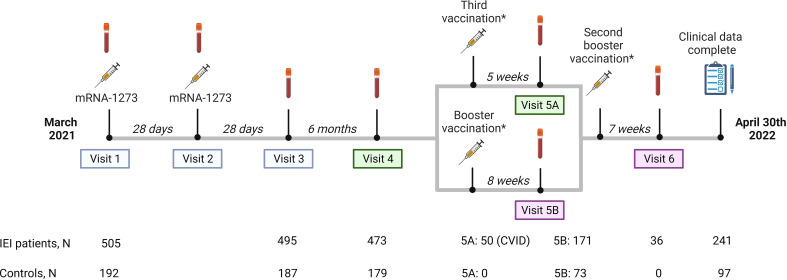
Methods: This study was embedded in a large prospective multicenter study investigating the immunogenicity of COVID-19 mRNA-based vaccines in IEI (VACOPID study). Blood samples were taken from 244 participants 8 weeks after booster vaccination. These participants included 171 IEI patients (X-linked agammaglobulinemia (XLA;N=11), combined immunodeficiency (CID;N=4), common variable immunodeficiency (CVID;N=45), isolated or undefined antibody deficiencies (N=108) and phagocyte defects (N=3)) and 73 controls. SARS-CoV-2-specific IgG titers, neutralizing antibodies, and T-cell responses were evaluated. One year after the start of the COVID-19 vaccination program, 334 study participants (239 IEI patients and 95 controls) completed a questionnaire to supplement their clinical data focusing on SARS-CoV-2 infections.

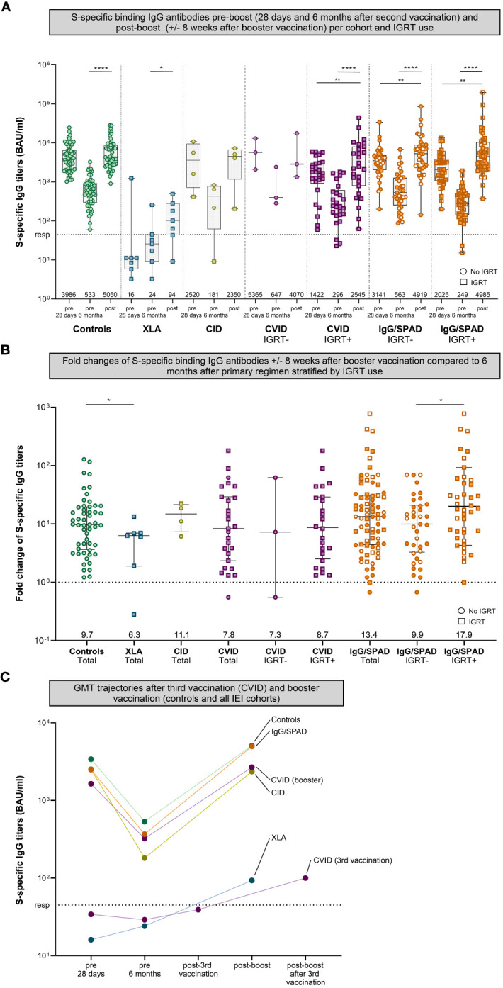
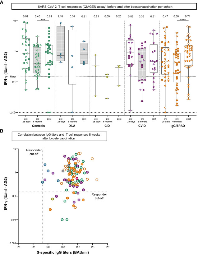
Results: After booster vaccination, S-specific IgG titers increased in all COVID-19 naive IEI cohorts and controls, when compared to titers at 6 months after the priming regimen. The fold-increases did not differ between controls and IEI cohorts. SARS-CoV-2-specific T-cell responses also increased equally in all cohorts after booster vaccination compared to 6 months after the priming regimen. Most SARS-CoV-2 infections during the study period occurred in the period when the Omicron variant had become dominant. The clinical course of these infections was mild, although IEI patients experienced more frequent fever and dyspnea compared to controls and their symptoms persisted longer.

Conclusion: Our study demonstrates that mRNA-based booster vaccination induces robust recall of memory B-cell and T-cell responses in most IEI patients. One-year clinical follow-up demonstrated that SARS-CoV-2 infections in IEI patients were mild. Given our results, we support booster campaigns with newer variant-specific COVID-19 booster vaccines to IEI patients with milder phenotypes.

Keywords: SARS-CoV-2; T-cell response; antibody response; booster vaccination; immunogenicity; inborn errors of immunity; mRNA-1273 COVID-19 vaccine; primary immunodeficiency disorders.


Conflict of interest statement

JP received a grant from GlaxoSmithKline for an improvement of clinical care project and received support from Prothva Biosolutions for attending meetings and cover of travel expenses. JP participates in an Advisory Board for Janssen. FV received a grant from ZonMW for a study on lanadelumab in COVID-19, and consulting fees from GSK made to his department. VD received consulting fees from GlaxoSmithKline, Pharming NV for Advisory board meetings and honoraria for lectures from Takeda Pharmaceutical Company, Kedrion, AstraZeneca. The remaining authors declare that the research was conducted in the absence of any commercial or financial relationships that could be construed as a potential conflict of interest. The author(s) declared that they were an editorial board member of Frontiers, at the time of submission. This had no impact on the peer review process and the final decision.

Figures

Figure 1
Figure 1
Study design. Study design including study visits, number of participants per study visit and median intervals between study visits or between vaccination and study visits. Study visit 5 is divided into part A and B. Part A are participants receiving a third vaccination. In our study, these participants are CVID patients who use immunosuppressive drugs, or in specific individual cases when a medical specialist had reasonable arguments to make an exception to the aforementioned indications, based on proven or assumed non-response. Part B are participants who received a regular booster vaccination. Results of study visits 1, 2 and 3, 4 and 5A were published previously (blue and green panels). Results of study visit 5B and 6 (pink panels) are included in this study.*The mRNA-1273 (Moderna)- and the BNT162b2 (Pfizer) vaccines were used as third vaccination or booster vaccination based on local availability and personal preferences and were administered at public vaccination sites.
Figure 2
Figure 2
S-specific IgG pre- (28 days and six months after second vaccination) and post-boost (+/- 8 weeks after booster vaccination). (A) S-specific IgG measured by Luminex for controls and different cohorts of inborn errors of immunity (IEI) patients 28 days after second vaccination, six months after the second vaccination, and eight weeks after booster vaccination. The number of participants per cohort correspond to Table 1 . The CVID and IgG cohort were stratified based on IGRT use. Results are expressed in binding antibody units per milliliter (BAU/mL). The dotted line is the pre-defined responder cut-off (resp). Data in panel A are presented in box-and-whisker plots. The horizontal lines of the box-and-whisker plots indicate the median, the bounds of the boxes indicate the interquartile range, and the whiskers indicate the range. All datapoints are shown. The numbers below the box-and-whisker plots indicate the geometric mean titers (GMT) per time point. Participants not using immunoglobulin replacement therapy (IGRT) are shown as circles, participants using IGRT are shown as squares. IgG titers were compared per cohort using the Wilcoxon paired signed rank test. The SPAD cohort is indicated with white symbols (with orange borders) while the IgG cohort is indicated with orange symbols. (B) Fold changes of IgG antibodies post-boost (+/- 8 weeks after booster vaccination) compared to 6 months after primary regimen of each cohort in total plus the fold changes the CVID and IgG/SPAD cohort stratified by IGRT use. Data in panel B are presented in scatter dot plots. The horizontal lines indicate the median, the whiskers indicate the interquartile range. All data points are shown. The dashed line represents a fold change of 1, where the titer at 6 months after primary regimen is equal to the titer after booster. All data points above the dashed line represents a fold increase, all data points below the dashed line a fold decrease. The numbers below indicate the median fold change. Fold changes were compared per cohort using the Wilcoxon rank-sum test. (C) Trajectories of the GMTs after third vaccination (CVID) and booster vaccination (all cohorts). The results of CVID participants receiving a third vaccination have also been published previously. A subset of these CVID participants received a booster after their third vaccination (fourth dose in total, named post-boost after 3rd vaccination on the x-axis). The results after booster vaccination of the CVID cohort and the other cohorts were computed only on those who donated blood after booster vaccination. Color coding is the same in all figures. S, Spike; XLA, X-linked agammaglobulinemia; CID, Combined Immunodeficiency; CVID, Common Variable Immunodeficiency; IgG, Isolated IgG subclass deficiency ± IgA deficiency; SPAD, Specific polysaccharide antibody deficiency; * = P<.05, ** = P<.01, *** = P<.001, **** = P<.0001.
Figure 3
Figure 3
SARS-CoV-2-specific T-cell responses pre-boost (28 days and six months after second vaccination) and post-boost (+/- 8 weeks after booster vaccination). (A) SARS-CoV-2-specific T-cell responses measured by an interferon γ (IFN-γ) release assay (QIAGEN) after stimulation of whole blood 28 days and six months after second vaccination and eight weeks after booster vaccination. Lower limit of detection (LLOD) is.01 IU/ml and responder cut off (resp) is.15 IU/ml. Results are expressed as international units/milliliter (IU/mL). The dotted line is the pre-defined responder cut-off (resp). Data is presented in box-and-whisker plots. The horizontal lines of the box-and-whisker plots indicate the median, the bounds of the boxes indicate the interquartile range, and the whiskers indicate the range. All datapoints are shown. The numbers above the box-and-whisker plots indicate the geometric mean titer (GMT). Within each cohort, IFN-γ levels at 28 days and six months were compared using Wilcoxon paired signed rank test. The SPAD cohort is indicated with white symbols while the IgG cohort is indicated with orange symbols. (B) Correlation between IgG titers and T-cell responses 8 weeks after booster vaccination. The dotted horizontal line is the responder cut-off of the QIAGEN interferon-gamma release assay (0.15 IU/mL). The dotted vertical line is the responder cut-off of the Luminex assay (44.8 BAU/ml). Color coding is the same in all figures. XLA, X-linked agammaglobulinemia; CID, Combined Immunodeficiency; CVID, Common Variable Immunodeficiency; IgG, Isolated IgG subclass deficiency ± IgA deficiency; SPAD, Specific polysaccharide antibody deficiency; *** = P<.001, **** = P<.0001.
Figure 4
Figure 4
SARS-CoV-2-specific IgG in CVID participants. (A) S-specific IgG measured by Luminex for CVID patients that received a third dose and a (fourth) booster dose after the initial regimen of 2 mRNA-1273 COVID-19 vaccines. S-specific IgG was measured 28 days after second vaccination, six months after the second vaccination, five weeks after third vaccination and eight weeks after booster vaccination. (B) S-specific IgG measured by Luminex for CVID patients after a third dose of a mRNA COVID-19 vaccine (either a third vaccination of a booster dose) stratified by the presence of non-infectious complications. The following non-infectious complications were defined: Autoimmune cytopenia, organ specific autoimmunity, systemic autoimmunity, enteropathy, malignancy, lymphoproliferative diseases, granulomatous lymphocytic interstitial lung disease (GLILD), and other granulomatous diseases. (A, B) Results are expressed in binding antibody units per milliliter (BAU/mL). The dotted line is the pre-defined responder cut-off. Data in panels A and B are presented in box-and-whisker plots. The horizontal lines of the box-and-whisker plots indicate the median, the bounds of the boxes indicate the interquartile range, and the whiskers indicate the range. All datapoints are shown. The numbers below the box-and-whisker plots indicate the geometric mean titer (GMT) per timepoint. IgG titers were compared per cohort using the Wilcoxon paired signed rank test. CVID, Common Variable Immunodeficiency; *** = P<.001, **** = P<.0001; ns = not significant.

Similar articles

Cited by

References

    1. van Leeuwen LPM, GeurtsvanKessel CH, Ellerbroek PM, de Bree GJ, Potjewijd J, Rutgers A, et al. . Immunogenicity of the mRNA-1273 COVID-19 vaccine in adult patients with inborn errors of immunity. J Allergy Clin Immunol. (2022) 149(6):1949–57. doi: 10.2139/ssrn.4001461 - DOI - PMC - PubMed
    1. Shields AM, Faustini SE, Hill HJ, Al-Taei S, Tanner C, Ashford F, et al. . SARS-CoV-2 vaccine responses in individuals with antibody deficiency: findings from the COV-AD study. J Clin Immunol. (2022) 42:923–34. doi: 10.1007/s10875-022-01231-7 - DOI - PMC - PubMed
    1. Durkee-Shock JR, Keller MD. Immunizing the imperfect immune system: Coronavirus disease 2019 vaccination in patients with inborn errors of immunity. Ann Allergy Asthma Immunol. (2022) 129(5):562–71. doi: 10.1016/j.anai.2022.06.009 - DOI - PMC - PubMed
    1. Ponsford MJ, Evans K, Carne EM, Jolles S, Immunodeficiency Centre for W. Division of Population M . COVID-19 vaccine uptake and efficacy in a national immunodeficiency cohort. J Clin Immunol. (2022) 42:728–31. doi: 10.1007/s10875-022-01223-7 - DOI - PMC - PubMed
    1. Delmonte OM, Bergerson JRE, Burbelo PD, Durkee-Shock JR, Dobbs K, Bosticardo M, et al. . Antibody responses to the SARS-CoV-2 vaccine in individuals with various inborn errors of immunity. J Allergy Clin Immunol. (2021) 148(5):1192–7. doi: 10.1016/j.jaci.2021.08.016 - DOI - PMC - PubMed

Publication types

MeSH terms