. 2024 Apr 18:15:1361240.
doi: 10.3389/fimmu.2024.1361240. eCollection 2024.

Changes in the N-glycosylation of porcine immune globulin G during postnatal development

Affiliations

Changes in the N-glycosylation of porcine immune globulin G during postnatal development

Kristina Zlatina et al. Front Immunol. .

Abstract

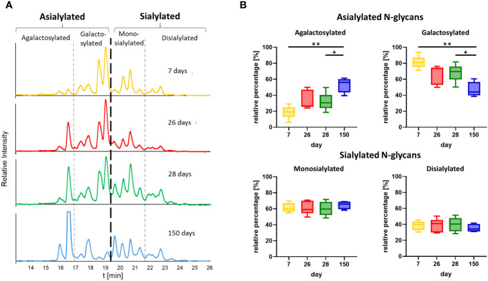
N-glycosylation influences the effectiveness of immune globulin G (IgG) and thus the immunological downstream responses of immune cells. This impact arises from the presence of N-glycans within the Fc region, which not only alters the conformation of IgG but also influences its steric hindrance. Consequently, these modifications affect the interaction between IgG and its binding partners within the immune system. Moreover, this posttranslational modification vary according to the physiological condition of each individual. In this study, we examined the N-glycosylation of IgG in pigs from birth to five months of age. Our analysis identified a total of 48 distinct N-glycan structures. Remarkably, we observed defined changes in the composition of these N-glycans during postnatal development. The presence of agalactosylated and sialylated structures increases in relation to the number of N-glycans terminated by galactose residues during the first months of life. This shift may indicate a transition from passively transferred antibodies from the colostrum of the sow to the active production of endogenous IgG by the pig's own immune system.

Keywords: N-glycan; antibody; glycosylation; ontogenesis; pig; porcine IgG.


Conflict of interest statement

The authors declare that the research was conducted in the absence of any commercial or financial relationships that could be construed as a potential conflict of interest. The author(s) declared that they were an editorial board member of Frontiers, at the time of submission. This had no impact on the peer review process and the final decision.

Figures

Figure 1
Figure 1
Structure of IgG with the conserved N-glycosylation site at asparagine (N) 297 in the Fc domain. Shown are representative N-glycan structures of human IgG grouped according to their potential to trigger pro- or anti-inflammatory effector functions of the immune system. Man, mannose; Gal, galactose; GlcNAc, N-acetylglucosamine; Fuc, fucose; Neu5Ac, N-acetylneuraminic acid. Created with BioRender.com.
Figure 2
Figure 2
Overview of sample collection and workflow. (A) Sample collection occurred on day 7 after birth, one day before (26) and one day after (28) weaning, as well as on the day of slaughtering on day 150. (B) The workflow displays the isolation of IgG from plasma by FPLC and the subsequent steps, including the verification of the IgG isolation by SDS-PAGE, the analysis of the glycan composition by GC-MS, the sialic acid quantification by RP-FD and the fluorescent analysis of N-glycans by HILIC-FD and HILIC-HESI-MS(/MS). Man, mannose; Gal, galactose; GlcNAc, N-Acetylglucosamine; Fuc, fucose; Neu5Ac, N-acetylneuraminic acid. Created with BioRender.com.
Figure 3
Figure 3
Verification of the purity of the isolated IgG from plasma, exemplary for one animal from all four time points. The isolated IgGs were separated by denaturing SDS-PAGE. Two micrograms of protein were separated and stained with Coomassie blue.
Figure 4
Figure 4
Glycan composition analysis during postnatal development. (A) N-glycan structures of IgGs and the respective proportions of the monosaccharides are shown. Created with BioRender.com. (B) Neutral monosaccharides were analyzed by GC-MS after hydrolysis and peracetylation. The proportions of GlcNAc (N-Acetylglucosamine), Man (mannose) and Gal (galactose) are displayed. Analyses that are more detailed are shown in Supplementary Figure 3 .
Figure 5
Figure 5
Sialylation status of IgGs during postnatal development. (A) Representative chromatograms of a standard containing fluorescently labelled Neu5Gc (N-glycolylneuraminic acid) and Neu5Ac (N-acetylneuraminic acid) and a sample are shown. (B) Analysis of sialic acids present on IgG by RP-chromatography. The amounts of Neu5Gc and Neu5Ac per 1 µg of IgG are shown. For the statistical analysis, mixed effect analysis and multiple comparison Tukey test were applied. Significant differences are given: * p < 0.1, ** p < 0.01, *** p < 0.001, and **** p < 0.0001.
Figure 6
Figure 6
N-glycan analysis of IgGs during postnatal development. (A) Fluorescently labelled N-glycans were separated by HILIC. A representative chromatogram from one animal at the age of 26 days is displayed. The peaks detected during the HILIC-FD analysis were fractionated and analyzed by UPLC-HESI-MS(/MS), and the identified glycans were assigned. The division of the chromatogram into a, mono- and disialylated structures confirmed the different retention times of the N-glycans. The chemical structures of Neu5Ac and Neu5Gc are also displayed. (B) All detected and assumed N-glycan structures based on the UPLC-HESI-MS(/MS)analysis. To depict the glycans, the Symbol Nomenclature For Glycans (SNFG) was used, and for their description, the Oxford Notation was selected. Man, mannose; Gal, galactose; GlcNAc, N-acetylglucosamine; Fuc, fucose; Neu5Ac, N-acetylneuraminic acid; Neu5Gc, N-glycolylneuraminic acid. Corresponding MS-data can be found in Supplementary Figure 1 .
Figure 7
Figure 7
Time point comparison of N-glycan composition. (A) Representative chromatograms of the N-glycans from one animal at all four time points are shown. The chromatograms are divided into peaks corresponding to asialylated and sialylated N-glycans. The asialylated N-glycans are additionally divided into agalactosylated and galactosylated N-glycans and the sialylated in mono- and disialylated N-glycans. The chromatogram at the age of 28 days corresponds to the chromatogram in Figure 6A . The chromatograms of all further animals can be found in Supplementary Figure 2 . (B) The peak areas of the agalactosylated, galactosylated, mono- and disialylated structures (please see Supplementary Figure 2 for the chromatograms) were determined and the ratios of the asialylated (agalactosylated/galactosylated) and sialylated (monosialylated/disialylated) N-glycans were calculated respectively. Box & Whisker plots (median; min to max) are shown. For the statistical analysis, mixed effect analysis and multiple comparison Tukey test were applied. Significant differences are given: * p < 0.1, ** p < 0.01.

Similar articles

References

    1. Arnold JN, Wormald MR, Sim RB, Rudd PM, Dwek RA. The impact of glycosylation on the biological function and structure of human immunoglobulins. Annu Rev Immunol. (2007) 25:21–50. doi: 10.1146/annurev.immunol.25.022106.141702 - DOI - PubMed
    1. Niwa R, Natsume A, Uehara A, Wakitani M, Iida S, Uchida K, et al. . IgG subclass-independent improvement of antibody-dependent cellular cytotoxicity by fucose removal from Asn297-linked oligosaccharides. J Immunol Methods. (2005) 306:151–60. doi: 10.1016/j.jim.2005.08.009 - DOI - PubMed
    1. Chung S, Quarmby V, Gao X, Ying Y, Lin L, Reed C, et al. . Quantitative evaluation of fucose reducing effects in a humanized antibody on Fcγ receptor binding and antibody-dependent cell-mediated cytotoxicity activities. MAbs. (2012) 4:326–40. doi: 10.4161/mabs.19941 - DOI - PMC - PubMed
    1. Shields RL, Lai J, Keck R, O'Connell LY, Hong K, Meng YG, et al. . Lack of fucose on human IgG1 N-linked oligosaccharide improves binding to human Fcgamma RIII and antibody-dependent cellular toxicity. J Biol Chem. (2002) 277:26733–40. doi: 10.1074/jbc.M202069200 - DOI - PubMed
    1. Dekkers G, Treffers L, Plomp R, Bentlage AE, de Boer M, Koeleman CA, et al. . Decoding the human immunoglobulin G-glycan repertoire reveals a spectrum of fc-receptor- and complement-mediated-effector activities. Front Immunol. (2017) 8:877. doi: 10.3389/fimmu.2017.00877 - DOI - PMC - PubMed

Publication types

LinkOut - more resources