. 2024 Apr 18:14:1382228.
doi: 10.3389/fcimb.2024.1382228. eCollection 2024.

Tissue-specific localization of tick-borne pathogens in ticks collected from camels in Kenya: insights into vector competence

Affiliations

Tissue-specific localization of tick-borne pathogens in ticks collected from camels in Kenya: insights into vector competence

Rua Khogali et al. Front Cell Infect Microbiol. .

Abstract

Background: Tick-borne pathogen (TBP) surveillance studies often use whole-tick homogenates when inferring tick-pathogen associations. However, localized TBP infections within tick tissues (saliva, hemolymph, salivary glands, and midgut) can inform pathogen transmission mechanisms and are key to disentangling pathogen detection from vector competence.

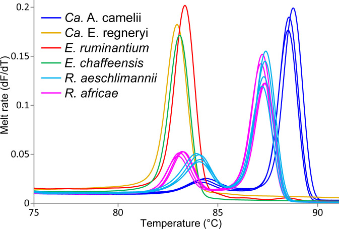
Methods: We screened 278 camel blood samples and 504 tick tissue samples derived from 126 camel ticks sampled in two Kenyan counties (Laikipia and Marsabit) for Anaplasma, Ehrlichia, Coxiella, Rickettsia, Theileria, and Babesia by PCR-HRM analysis.

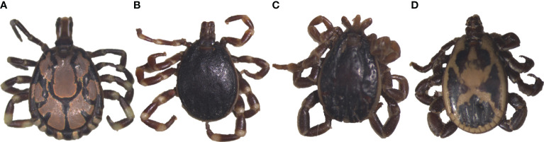
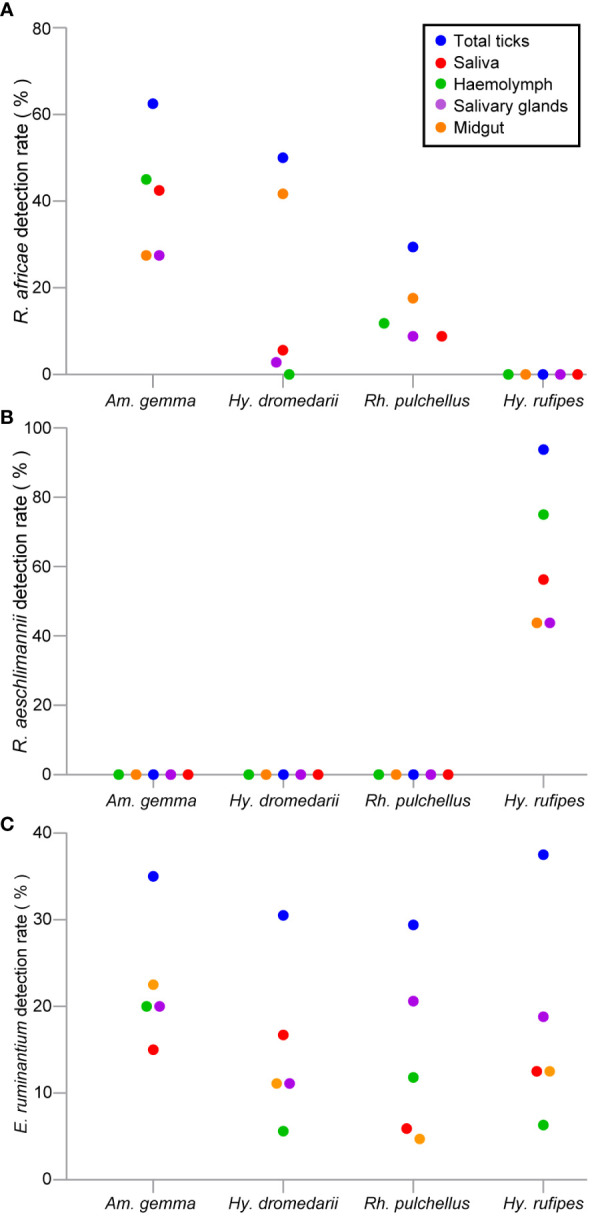
Results: Candidatus Anaplasma camelii infections were common in camels (91%), but absent in all samples from Rhipicephalus pulchellus, Amblyomma gemma, Hyalomma dromedarii, and Hyalomma rufipes ticks. We detected Ehrlichia ruminantium in all tissues of the four tick species, but Rickettsia aeschlimannii was only found in Hy. rufipes (all tissues). Rickettsia africae was highest in Am. gemma (62.5%), mainly in the hemolymph (45%) and less frequently in the midgut (27.5%) and lowest in Rh. pulchellus (29.4%), where midgut and hemolymph detection rates were 17.6% and 11.8%, respectively. Similarly, in Hy. dromedarii, R. africae was mainly detected in the midgut (41.7%) but was absent in the hemolymph. Rickettsia africae was not detected in Hy. rufipes. No Coxiella, Theileria, or Babesia spp. were detected in this study.

Conclusions: The tissue-specific localization of R. africae, found mainly in the hemolymph of Am. gemma, is congruent with the role of this tick species as its transmission vector. Thus, occurrence of TBPs in the hemolymph could serve as a predictor of vector competence of TBP transmission, especially in comparison to detection rates in the midgut, from which they must cross tissue barriers to effectively replicate and disseminate across tick tissues. Further studies should focus on exploring the distribution of TBPs within tick tissues to enhance knowledge of TBP epidemiology and to distinguish competent vectors from dead-end hosts.

Keywords: Amblyomma gemma; Ehrlichia; Hyalomma dromedarii; Hyalomma rufipes; Rhipicephalus pulchellus; Rickettsia; dromedary camels; tick tissues.


Conflict of interest statement

The authors declare that the research was conducted in the absence of any commercial or financial relationships that could be construed as a potential conflict of interest.

Figures

Figure 1
Figure 1
Maps showing the sampling sites where ticks and blood were collected from camels in Laikipia and Marsabit counties. Maps were generated using the open-source software, QGIS v.3.28.4.
Figure 2
Figure 2
Images of representative adult male ticks collected from camels in Laikipia and Marsabit counites. (A) Amblyomma gemma, (B) Hyalomma rufipes, (C) Hyalomma dromedarii, and (D) Rhipicephalus pulchellus. The images were captured using a Stemi 2000-C microscope (Zeiss, Oberkochen, Germany), a digital microscope, connected to an Axio-cam ERc 5s camera (Zeiss).
Figure 3
Figure 3
Maximum likelihood phylogenies of representative gene sequences amplified from ticks collected from camels in Laikipia and Marsabit counties. (A) tick CO1, (B) tick16S rRNA, and (C) tick 12S rRNA gene sequences. The study sequences, along with their respective GenBank accessions, are highlighted in bold. The bootstrap values at the nodes are the indicating percentage agreement from 1000 replicates. The branch length scale represents the substitution per site. Trees are rooted to outgroup sequences indicated in brackets.
Figure 4
Figure 4
Melt rate profiles of representative tick-borne pathogen PCR products amplified from camel blood and tick tissues. Melt rates are represented as a change in fluorescence with increasing temperature (dF/dT).
Figure 5
Figure 5
Maximum likelihood phylogenies of representative gene sequences amplified from tick-borne pathogens in tick tissues of camel ticks collected in Laikipia and Marasbit counties. (A) Anaplasamataceae 16S rRNA, (B) Ehrlichia 16S rRNA, and (C) Rickettsia ompB gene sequences. The study sequences, along with their respective GenBank accessions, are highlighted in bold. The bootstrap values at the nodes are the indicating percentage agreement from 1000 replicates. The branch length scale represents the substitution per site. Trees are rooted to outgroup sequences (indicated in brackets).
Figure 6
Figure 6
Scattered dot plots showing TBP detection rates (%) in tick tissues across different tick species. (A) R. africae, (B) R. aeschlimannii, and (C) E. ruminantium. All tick species were collected from camels in Marsabit and Laikipia counties.
Figure 7
Figure 7
Pearson correlation matrix heat map summarizing correlations of pathogen, R. africae, R. aeschlimannii, and E. ruminantium, occurrence and tissue type of infected individuals for different tick species. The different colors represent Pearson correlation coefficients, and the different circles sizes represent the P-value (*P < 0.05, **P < 0.01, ***P < 0.001). SL, saliva: HL, hemolymph; SG, salivary glands; MG, midgut.

Similar articles

Cited by

References

    1. Alanazi A. D., Nguyen V. L., Alyousif M. S., Manoj R. R. S., Alouffi A. S., Donato R., et al. . (2020). Ticks and associated pathogens in camels (Camelus dromedarius) from Riyadh Province, Saudi Arabia. Parasitol. Vectors. 13, 110. doi: 10.1186/s13071-020-3973-y - DOI - PMC - PubMed
    1. Altschul S. F., Gish W., Miller W., Myers E. W., Lipman D. J. (1990). Basic local alignment search tool. J. Mol. Biol. 215, 403–410. doi: 10.1016/S0022-2836(05)80360-2 - DOI - PubMed
    1. Bargul J., Kidambasi K., Getahun M., Villinger J., Copeland R., Muema J., et al. . (2021). Transmission of ‘Candidatus Anaplasma camelii’ to laboratory animals by camel-specific keds, Hippobosca camelina . PloS Negl. Trop. Dis. 15, e0009671. doi: 10.1371/journal.pntd.0009671 - DOI - PMC - PubMed
    1. Bastos A. D., Mohammed O. B., Bennett N. C., Petevinos C., Alagaili A. N. (2015). Molecular detection of novel Anaplasmataceae closely related to Anaplasma platys and Ehrlichia canis in the dromedary camel (Camelus dromedarius). Vet. Microbiol. 179, 310–314. doi: 10.1016/j.vetmic.2015.06.001 - DOI - PubMed
    1. Ben Said M., Belkahia H., Sayahi L., Aloui M., Jemli M. H., Hadj Mohamed B., et al. . (2013). First serological study of the prevalence of Anaplasma phagocytophilum in dromedary (Camelus dromedarius) in Tunisia. Bull. Soc Pathol. Exot. 107, 1–6. doi: 10.1186/s13071-020-3973-y - DOI - PubMed

Publication types

MeSH terms