. 2024 Jan-Mar;16(1):40-47.
doi: 10.32607/actanaturae.27338.

The Effect of Calcium Ions on the Electrophysiological Properties of Single ANO6 Channels

Affiliations

The Effect of Calcium Ions on the Electrophysiological Properties of Single ANO6 Channels

D O Kolesnikov et al. Acta Naturae. 2024 Jan-Mar.

Abstract

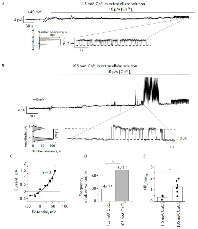
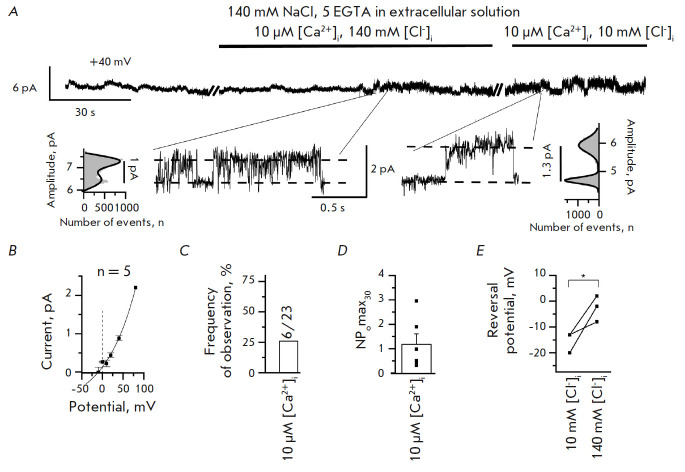
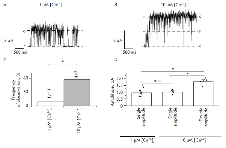
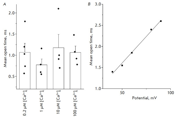
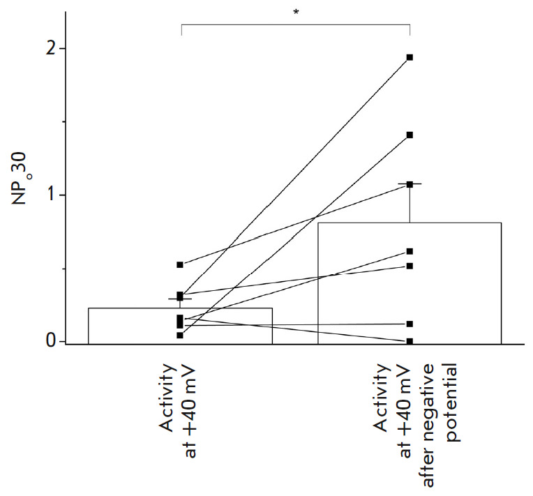
Proteins belonging to the anoctamin (ANO) family form calcium-activated chloride channels (CaCCs). The most unusual member of this family, ANO6 (TMEM16F), simultaneously exhibits the functions of calcium-dependent scramblase and the ion channel. ANO6 affects the plasma membrane dynamics and phosphatidylserine transport; it is also involved in programmed cell death. The properties of ANO6 channels remain the subject of debate. In this study, we investigated the effect of variations in the intracellular and extracellular concentrations of calcium ions on the electrophysiological properties of endogenous ANO6 channels by recording single ANO6 channels. It has been demonstrated that (1) a high calcium concentration in an extracellular solution increases the activity of endogenous ANO6 channels, (2) the permeability of endogenous ANO6 channels for chloride ions is independent of the extracellular concentration of calcium ions, (3) that an increase in the intracellular calcium concentration leads to the activation of endogenous ANO6 channels with double amplitude, and (4) that the kinetics of the channel depend on the plasma membrane potential rather than the intracellular concentration of calcium ions. Our findings give grounds for proposing new mechanisms for the regulation of the ANO6 channel activity by calcium ions both at the inner and outer sides of the membrane.

Keywords: ANO6; TMEM16F; calcium-activated chloride channels; patch-clamp technique; recording currents through single channels.


Figures

Fig. 1
Fig. 1
Activity of endogenous ANO6 CaCCs induced by application of 10 μM [Ca2+]i to the intracellular side of the plasma membrane in the inside-out configuration in the presence of 1.5 mM or 105 mM Ca2+ in the extracellular solution. Representative fragments of the recordings are shown, with expanded current traces and corresponding amplitude histograms at the bottom. (A) A fragment of the current recording through endogenous ANO6 CaCCs activated by application of 10 μM [Ca2+]i with 1.5 mM Ca2+ in the extracellular solution. (B) A fragment of the current recording through endogenous ANO6 CaCCs activated by application of 10 μM [Ca2+]i with 105 mM Ca2+ in the extracellular solution. (C) The average current–voltage relationship of endogenous ANO6 CaCCs in the presence of 1.5 mM Ca2+ in the extracellular solution. (D) The frequency of experiment observations with endogenous ANO6 CaCCs activated by application of 10 μM [Ca2+]i with 1.5 mM or 105 mM Ca2+ in the extracellular solution (p < 0.05). (E) NPomax30 of endogenous ANO6 CaCCs activated by application of 10 μM [Ca2+]i in the extracellular solutions of 1.5 mM and 105 mM Ca2+ (p < 0.05)
Fig. 2
Fig. 2
Activity of endogenous ANO6 CaCCs in the absence of divalent ions in the external solution. (A) A fragment of current recording through endogenous ANO6 CaCCs in an external solution containing 140 mM NaCl, 5 mM EGTA. Representative fragments of the recordings are shown, with expanded current traces and corresponding amplitude histograms at the bottom. (B) The average current–voltage relationship of endogenous CaCCs in an external solution containing 140 mM NaCl, 5 mM EGTA and intracellular solution based on CsGlutamate. (C) Frequency of observation of endogenous ANO6 CaCCs at 140 mM NaCl, 5 mM EGTA in an external solution. (D) The open state probability of endogenous ANO6 CaCCs at 140 mM NaCl, 5 mM EGTA in an external solution. (E) A shift in the reversal potential of endogenous CaCCs when one replaces an intracellular CsGlutamate solution with CsCl at 140 mM NaCl, 5 mM EGTA in the extracellular solution
Fig. 3
Fig. 3
Amplitude of endogenous CaCC at 1 and 10 μM [Ca2+]i. The extracellular solution contained 105 mM CaCl2. The channels were activated by application of a solution with 1 or 10 μM [Ca2+]i. (A) A fragment of current recording through endogenous single-amplitude CaCC activated by application of 1 μM [Ca2+]i. c is the closed state of the channel; o is the open state of the channel. (B) A fragment of current recording through endogenous double amplitude CaCC activated by application of 10 μM [Ca2+]i. c is the closed state of the channel; o is the open state of the channel; and s is the substate. (C) The frequency of observation of endogenous CaCC ANO6 with double amplitude activated by 1 μM [Ca2+]i (white) or 10 μM [Ca2+]i (gray), p < 0.05. (D) The amplitude of endogenous ANO6 channels activated by 1 or 10 μM [Ca2+]i (p < 0.05)
Fig. 4
Fig. 4
Effect of [Ca2+]i and the membrane potential on the open state lifetime of endogenous ANO6 channels in HEK293T cells. For the analysis, experiments were conducted in an inside-out configuration with 105 mM CaCl2 in the extracellular solution. (A) The average open state lifetime of endogenous CaCCs at +40 mV at various intracellular calcium concentrations (p > 0.05). (B) The dependence of the open state lifetime of endogenous CaCCs on the membrane potential at an intracellular calcium concentration of 100 μM (the data from a representative experiment are presented)
Fig. 5
Fig. 5
Activity of ANO6 channels at +40 mV before and after transient membrane potential switching to a negative value. The channels were pre-activated by adding 1 μM calcium to the intracellular side of the membrane. After the development of activity at +40 mV, the membrane potential was switched to negative and then returned to +40 mV

Similar articles

References

    1. Ehlen H.W.A., Chinenkova M., Moser M., Munter H.M., Krause Y., Gross S., Brachvogel B., Wuelling M., Kornak U., Vortkamp A., J. Bone Mineral Res. 2013;28(2):246–259. - PubMed
    1. Zhang Y., Le T., Grabau R., Mohseni Z., Kim H., Natale D.R., Feng L., Pan H., Yang H., Sci. Advanc. 2020;6:eaba0310. - PMC - PubMed
    1. Ousingsawat J., Wanitchakool P., Schreiber R., Wuelling M., Vortkamp A., Kunzelmann K.. J. Biol. Chem. 2015;290(10):6270–6280. - PMC - PubMed
    1. Yang H., Kim A., David T., Palmer D., Jin T., Tien J., Huang F., Cheng T., Coughlin S.R., Jan Y.N.. Cell. 2012;151(1):111–122. - PMC - PubMed
    1. Cabrita I., Benedetto R., Schreiber R., Kunzelmann K.. JCI Insight. 2019;4(15):e128414. - PMC - PubMed

LinkOut - more resources