. 2024 Apr 9;8(6):ziae050.
doi: 10.1093/jbmrpl/ziae050. eCollection 2024 Jun.

Loss-of-function OGFRL1 variants identified in autosomal recessive cherubism families

Affiliations

Loss-of-function OGFRL1 variants identified in autosomal recessive cherubism families

Mizuho Kittaka et al. JBMR Plus. .

Abstract

Cherubism (OMIM 118400) is a rare craniofacial disorder in children characterized by destructive jawbone expansion due to the growth of inflammatory fibrous lesions. Our previous studies have shown that gain-of-function mutations in SH3 domain-binding protein 2 (SH3BP2) are responsible for cherubism and that a knock-in mouse model for cherubism recapitulates the features of cherubism, such as increased osteoclast formation and jawbone destruction. To date, SH3BP2 is the only gene identified to be responsible for cherubism. Since not all patients clinically diagnosed with cherubism had mutations in SH3BP2, we hypothesized that there may be novel cherubism genes and that these genes may play a role in jawbone homeostasis. Here, using whole exome sequencing, we identified homozygous loss-of-function variants in the opioid growth factor receptor like 1 (OGFRL1) gene in 2 independent autosomal recessive cherubism families from Syria and India. The newly identified pathogenic homozygous variants were not reported in any variant databases, suggesting that OGFRL1 is a novel gene responsible for cherubism. Single cell analysis of mouse jawbone tissue revealed that Ogfrl1 is highly expressed in myeloid lineage cells. We generated OGFRL1 knockout mice and mice carrying the Syrian frameshift mutation to understand the in vivo role of OGFRL1. However, neither mouse model recapitulated human cherubism or the phenotypes exhibited by SH3BP2 cherubism mice under physiological and periodontitis conditions. Unlike bone marrow-derived M-CSF-dependent macrophages (BMMs) carrying the SH3BP2 cherubism mutation, BMMs lacking OGFRL1 or carrying the Syrian mutation showed no difference in TNF-ɑ mRNA induction by LPS or TNF-ɑ compared to WT BMMs. Osteoclast formation induced by RANKL was also comparable. These results suggest that the loss-of-function effects of OGFRL1 in humans differ from those in mice and highlight the fact that mice are not always an ideal model for studying rare craniofacial bone disorders.

Keywords: OGFRL1; autosomal recessive; cherubism; loss of function; mouse model; mutation; rare disease; whole exome sequencing.


Conflict of interest statement

All authors state that they have no conflicts of interest.

Figures

Graphical Abstract
Graphical Abstract
Figure 1
Figure 1
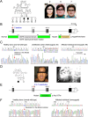
Loss-of-function mutations in the OGFRL1 gene identified in families affected with an autosomal recessive form of jawbone dysplasia; (A) left: the pedigree of a Syrian family affected with autosomal recessive jawbone dysplasia diagnosed with cherubism; right: facial appearances of individuals #2, 6, and 7; (B) top: the gene structure of OGFRL1; a red arrow indicates the location of the homozygous NM_024576.5:c.75del mutation (hereafter referred as c.75delC); bottom left: location of the putative OGFR conserved domain in OGFRL1; bottom right: frameshifted OGFRL1 protein (NP_078852.3:p.(Asp26ThrfsTer93)) due to the c.75delC mutation; (C) electropherograms of partial sequences of exon 1 of OGFRL1 showing the normal wt sequence and the heterozygous (het) or homozygous (hom) c.75delC mutation; Sanger sequencing confirmed that #1, 2, 3, 4, 5, 8 are heterozygous for the mutation, #6 and 7 are homozygous for the mutation, #9 has the WT sequence; (D) left: the pedigree of an Indian family affected with autosomal recessive jawbone dysplasia diagnosed as cherubism; middle: facial appearance of an affected individual; right: an X-ray image of the jawbone of the affected individual; white arrows indicate the expansile mandible; (E) top: the location of the homozygous NM_024576.5:c.337C > T mutation (hereafter referred as c.337C > T) in OGFRL1 found in the Indian family (red arrow); this gene mutation generated a truncated OGFRL1 protein (NP_078852.3:p.(Arg113Ter), hereafter referred as p.Arg113Ter) lacking the OGFR conserved domain; bottom: truncated OGFRL1 protein due to p.Arg113Ter mutation; (F) electropherograms of partial sequences of exon 3 of OGFRL1 showing the WT sequence and the homozygous c.337C > T mutation; (A, D) filled box = affected male; filled circle = affected female; open box = unaffected male; open circle = unaffected female; (B, E) filled boxes = exons coding for OGFRL1 protein; open boxes = 5’ and 3’ UTRs.
Figure 2
Figure 2
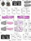
OGFRL1 knockout mice fail to recapitulate human cherubism; (A) facial appearance of OGFRL1-deficient male mice at 12 wk old; (B) two-dimensional microCT images of alveolar bone at the maxillary second molar; (C) microCT analysis of the alveolar bone underneath the maxillary second molar and the CEJ-ABC distance of the maxillary second molar; n = 11, 11, 14, 9, 11, 11, 14, 9, 11, 11, 14, 9 from left to right; (D) three-dimensional microCT images of the maxilla and 3 molars; (E) H&E staining images of the maxilla; (F) three-dimensional microCT images of the calvaria and H&E staining images of calvarial tissues; (G) TRAP staining of the alveolar bone underneath the maxillary second molar and histomorphometric analysis for osteoclasts; n = 5, 5, 5, 7 from left to right for both N.Oc/BS and Oc.S/BS; (H) left: two-dimensional microCT images of the alveolar bone at the maxillary second molar after ligature placement; right: microCT analysis of BV underneath the maxillary second molar with or without ligature placement and the percentage of alveolar bone loss underneath the maxillary second molar after ligature placement; n = 5, 6, 5, 6, 5, 12, 5, 12, 5, 6, 5, 12 from left to right; (E, F, G) bar = 500 μm. *P < .05 with Tukey–Kramer post hoc test; NS = not significant; data are presented with box and whisker plots.
Figure 3
Figure 3
OGFRL1 mice carrying a mutation equivalent to that in the Syrian family fail to recapitulate human cherubism; (A) facial appearance of OGFRL1 knock-in male mice at 12 wk old; (B) two-dimensional microCT images of alveolar bone at the maxillary second molar; (C) microCT analysis of the alveolar bone underneath the maxillary second molar and the CEJ-ABC distance of the maxillary second molar; n = 8, 10, 9, 9, 9, 13, 11, 10, 9, 13, 11, 10 from left to right; (D) three-dimensional microCT images of the maxilla and three molars; (E) H&E staining images of the maxilla; (F) three-dimensional microCT images of the calvaria and H&E staining images of calvarial tissues; (G) TRAP staining of the alveolar bone underneath the maxillary second molar and histomorphometric analysis for osteoclasts; n = 8, 8, 8, 8 from left to right for both N.Oc/BS and Oc.S/BS; (H) left: two-dimensional microCT images of the alveolar bone at the maxillary second molar after ligature placement; right: microCT analysis of BV underneath the maxillary second molar with or without ligature placement and the percentage of alveolar bone loss underneath the maxillary second molar after ligature placement; n = 8, 10, 7, 9, 9, 9, 10, 9, 8, 9, 8, 9 from left to right; (C, G, H) Del-c = Del-c/Del-c homozygote. (E, F, G) bar = 500 μm. *P < .05 with Tukey–Kramer post hoc test; NS = not significant; data are presented with box and whisker plots.

References

    1. Papadaki ME, Lietman SA, Levine MA, Olsen BR, Kaban LB, Reichenberger EJ. Cherubism: best clinical practice. Orphanet J Rare Dis. 2012;7(Suppl 1):S6. 10.1186/1750-1172-7-S1-S6 - DOI - PMC - PubMed
    1. Reichenberger EJ, Levine MA, Olsen BR, Papadaki ME, Lietman SA. The role of SH3BP2 in the pathophysiology of cherubism. Orphanet J Rare Dis. 2012;7(Suppl 1):S5. 10.1186/1750-1172-7-S1-S5 - DOI - PMC - PubMed
    1. Yoshitaka T, Mukai T, Kittaka M, et al. Enhanced TLR-MYD88 signaling stimulates autoinflammation in SH3BP2 cherubism mice and defines the etiology of cherubism. Cell Rep. 2014;8(6):1752–1766. 10.1016/j.celrep.2014.08.023 - DOI - PMC - PubMed
    1. Prod'Homme V, Boyer L, Dubois N, et al. Cherubism allele heterozygosity amplifies microbe-induced inflammatory responses in murine macrophages. J Clin Invest. 2015;125(4):1396–1400. 10.1172/JCI71081 - DOI - PMC - PubMed
    1. Ueki Y, Tiziani V, Santanna C, et al. Mutations in the gene encoding c-Abl-binding protein SH3BP2 cause cherubism. Nat Genet. 2001;28(2):125–126. 10.1038/88832 - DOI - PubMed