

Tailoring micellar nanocarriers for pemetrexed in breast cancer: design, fabrication and in vitro evaluation
- PMID: 38700294
- PMCID: PMC11418286
- DOI: 10.2217/nnm-2024-0013
Tailoring micellar nanocarriers for pemetrexed in breast cancer: design, fabrication and in vitro evaluation
Abstract
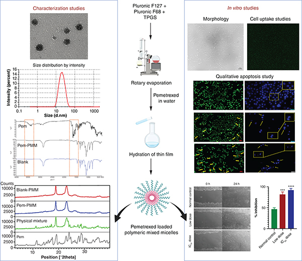
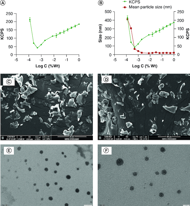
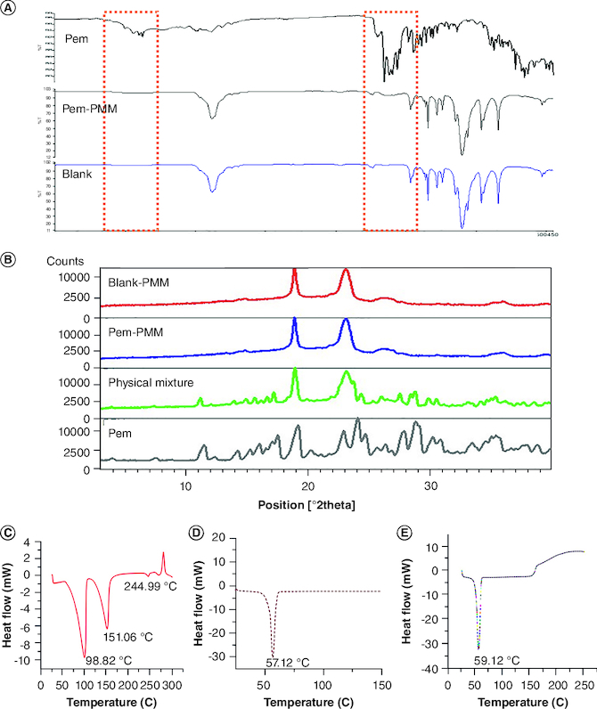
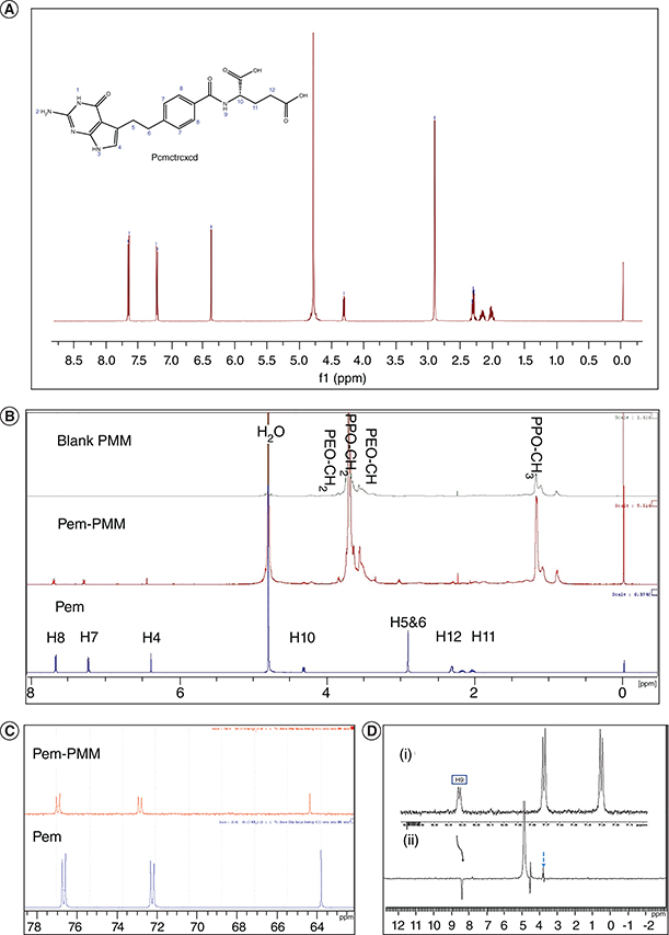
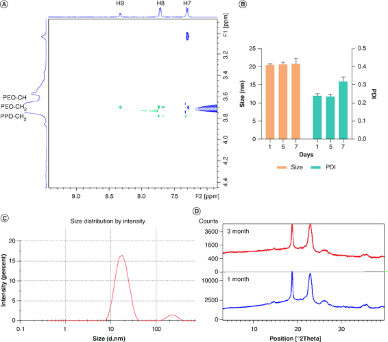
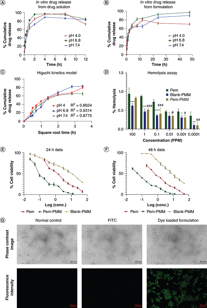
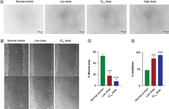
Aim: To investigate the pemetrexed encapsulated polymeric mixed micelles (PMMs) against breast cancer treatment.Methods: We meticulously optimized the formulation and conducted extensive characterizations, including photon correlation spectroscopy for micellization, advanced analytical techniques and in vitro cell line assessments.Results: The PMM exhibited favorable characteristics, with a spherical morphology, hydrodynamic particle size of 19.58 ± 0.89 nm, polydispersity index of 0.245 ± 0.1, and a surface charge of -9.70 ± 0.61 mV. Encapsulation efficiency and drug payload reached 96.16 ± 0.37% and 4.5 ± 0.32%, respectively. Cytotoxicity analysis indicated superior efficacy of the PMM over the drug solution.Conclusion: The PMM formulation exhibited controlled release of the drug, and demonstrated enhanced cytotoxicity against breast cancer cells, highlighting its therapeutic promise.
Keywords: TPGS; breast cancer; pemetrexed; pluronics; polymeric mixed micelles.
Plain language summary
[Box: see text].
Conflict of interest statement
The authors have no competing interests or relevant affiliations with any organization or entity with the subject matter or materials discussed in the manuscript. This includes employment, consultancies, honoraria, stock ownership or options, expert testimony, grants or patents received or pending, or royalties.
Figures

Similar articles
-
Enhancing breast cancer treatment: Comprehensive study of gefitinib-loaded poloxamer 407/TPGS mixed micelles through design, development, in-silico modelling, In-Vitro testing, and Ex-Vivo characterization.Int J Pharm. 2024 May 25;657:124109. doi: 10.1016/j.ijpharm.2024.124109. Epub 2024 Apr 16. Int J Pharm. 2024. PMID: 38626846
-
Celastrol-loaded polymeric mixed micelles shows improved antitumor efficacy in 4 T1 bearing xenograft mouse model through spatial targeting.Int J Pharm. 2024 Jun 25;659:124234. doi: 10.1016/j.ijpharm.2024.124234. Epub 2024 May 17. Int J Pharm. 2024. PMID: 38763310
-
Formulation and evaluation of mixed polymeric micelles of quercetin for treatment of breast, ovarian, and multidrug resistant cancers.Int J Nanomedicine. 2018 May 16;13:2869-2881. doi: 10.2147/IJN.S153094. eCollection 2018. Int J Nanomedicine. 2018. PMID: 29844670 Free PMC article.
-
Mixed micelles for encapsulation of doxorubicin with enhanced in vitro cytotoxicity on breast and ovarian cancer cell lines versus Doxil®.Biomed Pharmacother. 2017 Nov;95:894-903. doi: 10.1016/j.biopha.2017.09.006. Epub 2017 Sep 10. Biomed Pharmacother. 2017. PMID: 28903185
-
Benchmarking of pH-responsive mixed micelles for repurposed breast cancer therapy of ibrutinib with molecular modeling and pharmacokinetic insights.J Mater Chem B. 2025 Jun 11;13(23):6819-6842. doi: 10.1039/d5tb00419e. J Mater Chem B. 2025. PMID: 40401393
Cited by
-
The potential of nanoparticle-based siRNA delivery in breast cancer treatment.Nanomedicine (Lond). 2025 Mar;20(6):531-533. doi: 10.1080/17435889.2024.2440302. Epub 2024 Dec 10. Nanomedicine (Lond). 2025. PMID: 39655626 No abstract available.
References
-
- Kumari NU, Pardhi E, Chary PS, Mehra NK. Exploring contemporary breakthroughs in utilizing vesicular nanocarriers for breast cancer therapy. Ther. Deliv. 15(4), 279–303 (2024). - PubMed
-
- Dinakar YH, Rajana N, Kumari NU, Jain V, Mehra NK. Recent advances of multifunctional PLGA nanocarriers in the management of triple-negative breast cancer. AAPS PharmSciTech 24(8), 1–20 (2023). - PubMed
-
- Rajana N, Mounika A, Chary PSet al. . Multifunctional hybrid nanoparticles in diagnosis and therapy of breast cancer. J. Control. Rel. 352, 1024–1047 (2022). - PubMed
-
- Gridelli C, Maione P, Rossi Aet al. . Pemetrexed in advanced non-small-cell lung cancer. Expert Opin. Drug Saf. 10(2), 311–317 (2011). - PubMed
MeSH terms
Substances
LinkOut - more resources
Full Text Sources
Other Literature Sources
Medical