. 2024 Jul;13(19):e2304618.
doi: 10.1002/adhm.202304618. Epub 2024 May 17.

Prostate-Specific Membrane Antigen Targeted StarPEG Nanocarrier for Imaging and Therapy of Prostate Cancer

Affiliations

Prostate-Specific Membrane Antigen Targeted StarPEG Nanocarrier for Imaging and Therapy of Prostate Cancer

Niranjan Meher et al. Adv Healthc Mater. 2024 Jul.

Abstract

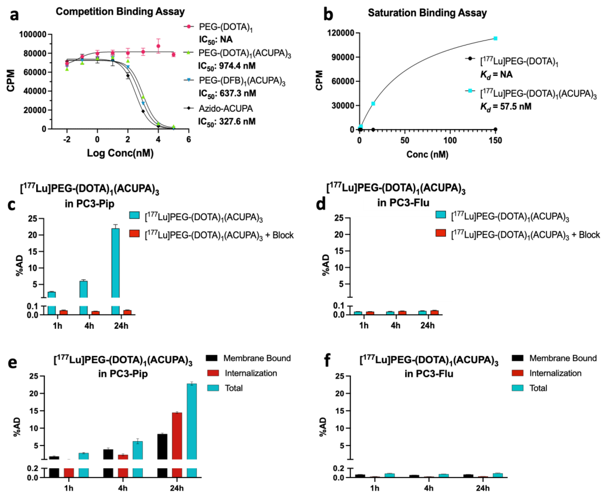
The tumor uptake of large non-targeted nanocarriers primarily occurs through passive extravasation, known as the enhanced permeability and retention (EPR) effect. Prior studies demonstrated improved tumor uptake and retention of 4-arm 40 kDa star polyethylene glycol (StarPEG) polymers for cancer imaging by adding prostate-specific membrane antigen (PSMA) targeting small molecule ligands. To test PSMA-targeted delivery and therapeutic efficacy, StarPEG nanodrugs with/without three copies of PSMA-targeting ligands, ACUPA, are designed and synthesized. For single-photon emission computed tomography (SPECT) imaging and therapy, each nanocarrier is labeled with 177Lu using DOTA radiometal chelator. The radiolabeled nanodrugs, [177Lu]PEG-(DOTA)1 and [177Lu]PEG-(DOTA)1(ACUPA)3, are evaluated in vitro and in vivo using PSMA+ PC3-Pip and/or PSMA- PC3-Flu cell lines, subcutaneous xenografts and disseminated metastatic models. The nanocarriers are efficiently radiolabeled with 177Lu with molar activities 10.8-15.8 MBq/nmol. Besides excellent in vitro PSMA binding affinity (kD = 51.7 nM), the targeted nanocarrier, [177Lu]PEG-(DOTA)1(ACUPA)3, demonstrated excellent in vivo SPECT imaging contrast with 21.3% ID/g PC3-Pip tumors uptake at 192 h. Single doses of 18.5 MBq [177Lu]PEG-(DOTA)1(ACUPA)3 showed complete resolution of the PC3-Pip xenografts observed up to 138 days. Along with PSMA-targeted excellent imaging contrast, these results demonstrated high treatment efficacy of [177Lu]PEG-(DOTA)1(ACUPA)3 for prostate cancer, with potential for clinical translation.

Keywords: enhanced permeability and retention; polymer nanocarriers; prostate cancer; prostate‐specific membrane antigen (PSMA); radioligand therapy; single photon excited computed tomography (SPECT) imaging.


Conflict of interest statement

D. Santi and G. Ashley, are employees of Prolynx Inc. The remaining authors declare no competing financial interest.

Competing interests

The authors declare no competing interests.

Figures

Figure 1.
Figure 1.
Graphical representation of PSMA-targeted μSPECT/CT imaging and therapy of prostate cancer using 177Lu-labeled StarPEG nanocarrier.
Figure 2.
Figure 2.
In vitro cell-binding assays with starPEG nanocarriers in PSMA+ PC3-Pip and PSMA− PC3-Flu cell lines demonstrate efficient cell binding and uptake of PSMA-targeted theranostic agents. (a) IC50 of nonradiolabeled theranostic StarPEGs, previously evaluated diagnostic nanocarrier PEG-(DFB)1(ACUPA)3 and Azido-ACUPA determined by [68Ga]PSMA-11-based in vitro competitive radioligand binding assay in PSMA+ PC3-Pip cells. (b) Kd measurement of 177Lu-labeled StarPEGs in the PSMA+ PC3-Pip cell line by a saturation binding assay. (c,d) Blocking assay of [177Lu]PEG-(DOTA)1(ACUPA)3 labeled starPEGs (5 nM) in PSMA+ PC3-Pip and PSMA− PC3-Flu cells using PSMA-2 (10 μM) as the blocking agent at 1 h, 4 h and 24h (%AD = percentage added dose). Blocking assays with the nontargeting [177Lu]PEG-(DOTA)1 have been presented in the Supporting Information (Figure S6). (e,f) Membrane-bound and internalization assay of the [177Lu]PEG-(DOTA)1(ACUPA)3 at 1 h, 4 h and 24 h in PSMA+ PC3-Pip and PSMA− PC3-Flu cells (%AD = percentage added dose). Membrane-bound and internalization assay with the nontargeting [177Lu]PEG-(DOTA)1 have been presented in the Supporting Information (Figure S7). Overall, the ACUPA conjugated StarPEG nanocarrier demonstrated excellent PSMA targeted binding affinity in PSMA+ PC3-Pip cells. (n=3, mean ± SD)
Figure 3.
Figure 3.
In vivo μSPECT/CT imaging demonstrates PSMA-targeted accumulation of StarPEG nanocarriers in PSMA+ PC3-Pip subcutaneous tumors. (a) Representation of experimental design for in vivo evaluation of the 177Lu-labeled starPEGs in mice bearing dual xenografts of PSMA+ PC3-Pip (left flank) and PSMA− PC3-Flu (right flank). (b) Maximum intensity projection (MIP) μSPECT/CT and coronal μSPECT/CT images obtained on day 24 h, 72 h, 144 h and 192 h post-injection of 177Lu-labeled starPEGs reveal high tumor accumulation of targeted nanocarriers in PSMA+ PC3-Pip. (c-d) Quantification of in vivo tumors’ accumulation by drawing ROIs on the respective tumors at 24 h, 72 h, 144 h and 192 h post-injection of 177Lu-labeled starPEGs. ROIs on the heart at respective imaging time points are presented in the Supporting Information (Figure S9). Overall, the in vivo μSPECT/CT imaging demonstrated excellent PSMA targeted tumor accumulation of the ACUPA conjugated StarPEG nanocarrier in PSMA+ PC3-Pip xenografts. (n=2, mean ± SD)
Figure 4.
Figure 4.
Ex vivo organ biodistribution of 177Lu-labeled starPEG nanocarriers. Organ biodistribution of (a) [177Lu]PEG-(DOTA)1 and (b) [177Lu]PEG-(DOTA)1(ACUPA)3 at 72 h and 192 h post-injection of the nanocarriers (n = 3, mean ± SD). The ratio of (c) PC3-Pip to PC3-Flu, (d) PC3-Pip to the muscle and (e) PC3-Pip to the blood of [177Lu]starPEGs at 72 h and 192 h post-injection of the nanocarriers (n=3, mean ± SD). The supporting information presents ex vivo organ biodistribution of [177Lu]starPEGs in % ID/Organ (Figure S10). Overall, the ex vivo organ biodistribution demonstrated excellent PSMA targeted tumor accumulation of the ACUPA conjugated StarPEG nanocarrier in PSMA+ PC3-Pip xenografts.
Figure 5.
Figure 5.
Autoradiography images of 20 μm tumor slices of PSMA+ PC3-Pip and PSMA− PC3-Flu tumors collected after 72 h and 192 h post-injection of [177Lu]StarPEG nanocarriers. Overall, the targeted nanocarrier demonstrated superior tissue penetration in PSMA+ PC3-Pip xenografts.
Figure 6.
Figure 6.
In vivo treatment study of [177Lu]PEG-(DOTA)1(ACUPA)3 in nude mice models bearing PSMA+ PC3-Pip tumors. (a) Representation of experimental design for in vivo treatment efficacy and toxicity of 177Lu-labeled starPEGs in mice models bearing PSMA+ PC3-Pip subcutaneous tumors. Tumor volume (b), body weight (c) and survival (d) of mice models bearing PC3-Pip tumors and treated with different doses (0, 4.6, 9.2 and 18.5, MBq) of either [177Lu]PEG-(DOTA)1(ACUPA)3 or 9.2 MBq of [177Lu]PSMA-617. Overall, a single 18.5 MBq dose of [177Lu]PEG-(DOTA)1(ACUPA)3 demonstrated highly efficient suppression of the tumor volume without any regrowth of tumors with 80% survival at 138 days post drug injection. (n=10, mean ± SD)
Figure 7.
Figure 7.
[177Lu]PEG-(DOTA)1(ACUPA)3 was effective in suppressing the growth of metastatic prostate cancer in the PC3-PiP intracardiac metastatic model. (a) Representation of experimental design for in vivo treatment study of the 177Lu-labeled starPEGs in mice bearing PSMA+ PC3-Pip metastatic tumors. (b) Multiple time points [68Ga]PSMA-11 μPET/CT imaging of the nude mice bearing PC3-Pip metastatic tumors before and after treatment of 9.2 MBq [177Lu]PEG-(DOTA)1(ACUPA)3 (n=4, mean ± SD). (c) Body weight measurement of the treated and control mice up to 51 days post-injection of 9.2 MBq [177Lu]PEG-(DOTA)1(ACUPA)3. (d) Overall survival of mice models bearing PC3-Pip metastatic tumors and treated with/without 9.2 MBq) of [177Lu]PEG-(DOTA)1(ACUPA)3. (e,f) [68Ga]PSMA-11 organ biodistribution of the nude mice bearing PC3-Pip metastatic tumors on 35 and 50 days post-treatment of 9.2 MBq [177Lu]PEG-(DOTA)1(ACUPA)3 (n=10, mean ± SD). Overall, a single 9.2 MBq dose of [177Lu]PEG-(DOTA)1(ACUPA)3 demonstrated highly efficient suppression of PC3-Pip metastatic tumors with 100% survival at 51 days post drug injection.
Scheme 1.
Scheme 1.
Synthetic routes to the StarPEG theranostic conjugates (a) PEG-(DOTA)1 and (b) PEG-(DOTA)1(ACUPA)3 evaluated in PSMA+ subcutaneous as well metastatic tumor models. Detailed synthetic protocols are provided in the method section.

Similar articles

References

    1. Wang H, He Z, Liu X, Huang Y, Hou J, Zhang W, Ding D, Small Structures 2022, 3, 2200036.
    1. Davis M, Bennett M, Thomas L, Bjorkman P, Proceedings of the National Academy of Sciences of the United States of America 2005, 102, 5981. - PMC - PubMed
    1. Journal of Nuclear Medicine 2022, 63, 13N.
    1. Pastorino S, Riondato M, Uccelli L, Giovacchini G, Giovannini E, Duce V, Ciarmiello A, Current Radiopharmaceuticals 2020, 13, 63. - PMC - PubMed
    2. Filippi L, Chiaravalloti A, Schillaci O, Bagni O, Expert Review of Anticancer Therapy 2020, 20, 823. - PubMed
    1. Price E, Orvig C, Chemical Society Reviews 2014, 43, 260. - PubMed
    2. Hofmann M, Maecke H, Borner A, Weckesser E, Schoffski P, Oei M, Schumacher J, Henze M, Heppeler A, Meyer G, Knapp W, European Journal of Nuclear Medicine 2001, 28, 1751. - PubMed
    3. Debela D, Muzazu S, Heraro K, Ndalama M, Mesele B, Haile D, Kitui S, Manyazewal T, Sage Open Medicine 2021, 9. - PMC - PubMed

MeSH terms