

Sensitivity and activation of endoplasmic reticulum stress response and apoptosis in the perinatal sheep heart
- PMID: 38700493
- PMCID: PMC11380940
- DOI: 10.1152/ajpheart.00043.2024
Sensitivity and activation of endoplasmic reticulum stress response and apoptosis in the perinatal sheep heart
Abstract
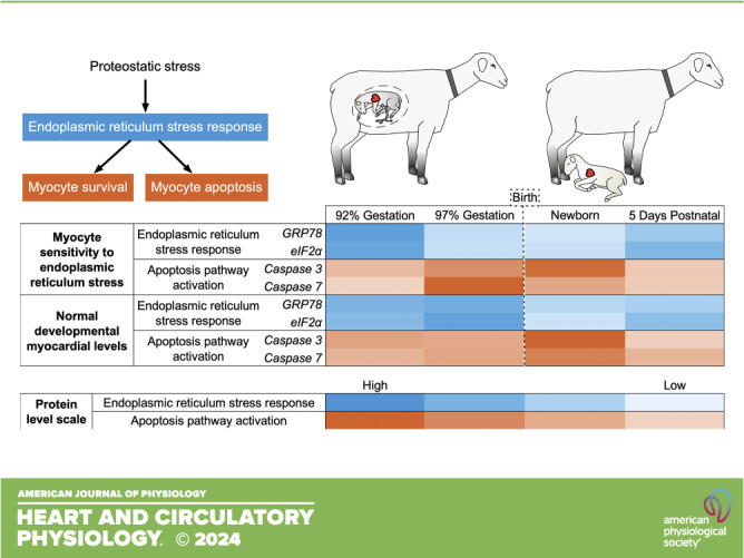
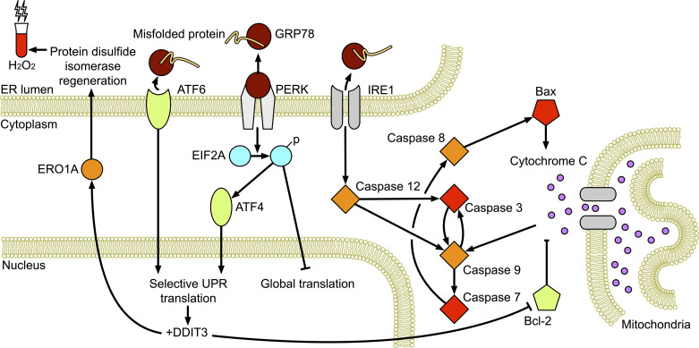
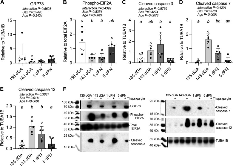
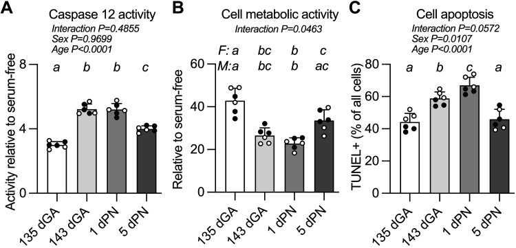
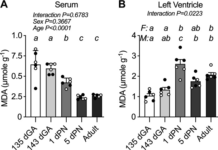
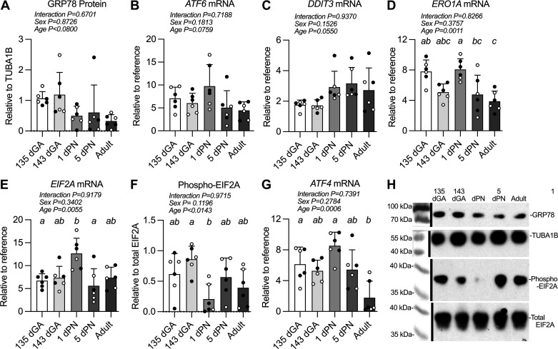
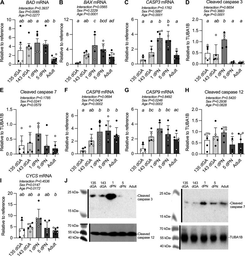
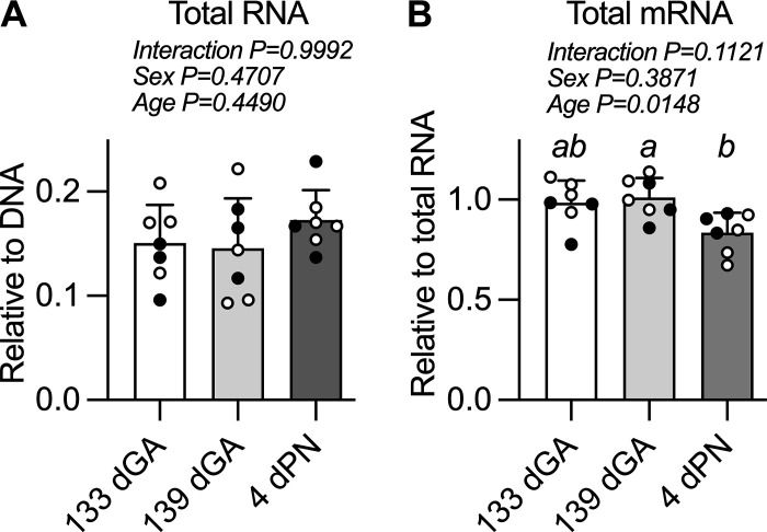
Although the unfolded protein response (UPR) contributes to survival by removing misfolded proteins, endoplasmic reticulum (ER) stress also activates proapoptotic pathways. Changed sensitivity to normal developmental stimuli may underlie observed cardiomyocyte apoptosis in the healthy perinatal heart. We determined in vitro sensitivity to thapsigargin in sheep cardiomyocytes from four perinatal ages. In utero cardiac activation of ER stress and apoptotic pathways was determined at these same ages. Thapsigargin-induced phosphorylation of eukaryotic initiation factor 2 (EIF2A) was decreased by 72% between 135 and 143 dGA (P = 0.0096) and remained low at 1 dPN (P = 0.0080). Conversely, thapsigargin-induced caspase cleavage was highest around the time of birth: cleaved caspase 3 was highest at 1 dPN (3.8-fold vs. 135 dGA, P = 0.0380; 7.8-fold vs. 5 dPN, P = 0.0118), cleaved caspase 7 and cleaved caspase 12 both increased between 135 and 143 dGA (25-fold and 6.9-fold respectively, both P < 0.0001) and remained elevated at 1 dPN. Induced apoptosis, measured by TdT-mediated dUTP nick-end labeling (TUNEL) assay, was highest around the time of birth (P < 0.0001). There were changes in myocardial ER stress pathway components in utero. Glucose (78 kDa)-regulated protein (GRP78) protein levels were high in the fetus and declined after birth (P < 0.0001). EIF2A phosphorylation was profoundly depressed at 1 dPN (vs. 143 dGA, P = 0.0113). In conclusion, there is dynamic regulation of ER proteostasis, ER stress, and apoptosis cascade in the perinatal heart. Apoptotic signaling is more readily activated in fetal cardiomyocytes near birth, leading to widespread caspase cleavage in the newborn heart. These pathways are important for the regulation of normal maturation in the healthy perinatal heart.NEW & NOTEWORTHY Cardiomyocyte apoptosis occurs even in the healthy, normally developing perinatal myocardium. As cardiomyocyte number is a critical contributor to heart health, the sensitivity of cardiomyocytes to endoplasmic reticulum stress leading to apoptosis is an important consideration. This study suggests that the heart has less robust protective mechanisms in response to endoplasmic reticulum stress immediately before and after birth, and that more cardiomyocyte death can be induced by stress in this period.
Keywords: cardiac; caspases; fetal development; myocardium; unfolded protein response.
Conflict of interest statement
No conflicts of interest, financial or otherwise, are declared by the authors.
Figures
Similar articles
-
Eukaryotic translation initiation factor 2A protects pancreatic beta cells during endoplasmic reticulum stress while rescuing global translation inhibition.Diabetologia. 2025 Aug;68(8):1735-1753. doi: 10.1007/s00125-025-06431-5. Epub 2025 Apr 30. Diabetologia. 2025. PMID: 40304759
-
[Crosstalk between activating transcription factor 6 and the inositol-requiring enzyme 1-X-box binding protein 1 pathway in oxygen-glucose deprivation/reoxygenation-injured HT22 cells].Zhonghua Wei Zhong Bing Ji Jiu Yi Xue. 2023 Mar;35(3):278-286. doi: 10.3760/cma.j.cn121430-20230228-00115. Zhonghua Wei Zhong Bing Ji Jiu Yi Xue. 2023. PMID: 36916341 Chinese.
-
Amplifying Endoplasmic Reticulum Stress With Adenosine Triphosphate-Coated Gold Nanoclusters: A Promising Approach for the Treatment of Vestibular Schwannoma.Otol Neurotol. 2025 Aug 1;46(7):e269-e277. doi: 10.1097/MAO.0000000000004403. Otol Neurotol. 2025. PMID: 40644638
-
Modulation of Endoplasmic Reticulum Stress in Experimental Anti-Cancer Therapy.Int J Mol Sci. 2025 Jul 3;26(13):6407. doi: 10.3390/ijms26136407. Int J Mol Sci. 2025. PMID: 40650182 Free PMC article. Review.
-
Fetal scalp stimulation for assessing fetal well-being during labour.Cochrane Database Syst Rev. 2023 Jan 10;1(1):CD013808. doi: 10.1002/14651858.CD013808.pub2. Cochrane Database Syst Rev. 2023. PMID: 36625680 Free PMC article.
References
-
- Adler CP, Costabel U. Cell number in human heart in atrophy, hypertrophy, and under the influence of cytostatics. Recent Adv Stud Cardiac Struct Metab 6: 343–355, 1975. - PubMed
Publication types
MeSH terms
Substances
Grants and funding
LinkOut - more resources
Full Text Sources
Research Materials
Miscellaneous