. 2024 Jul;62(7):555-568.
doi: 10.1007/s12275-024-00133-0. Epub 2024 May 3.

Genetically Engineered CLDN18.2 CAR-T Cells Expressing Synthetic PD1/CD28 Fusion Receptors Produced Using a Lentiviral Vector

Affiliations

Genetically Engineered CLDN18.2 CAR-T Cells Expressing Synthetic PD1/CD28 Fusion Receptors Produced Using a Lentiviral Vector

Heon Ju Lee et al. J Microbiol. 2024 Jul.

Abstract

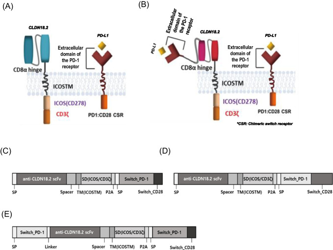
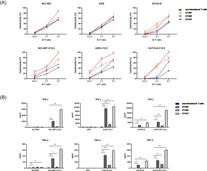
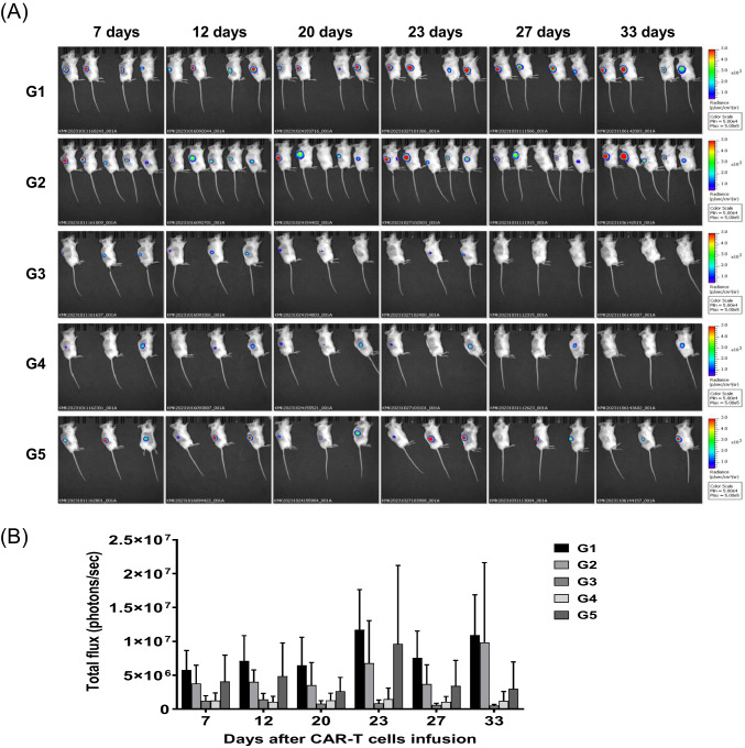
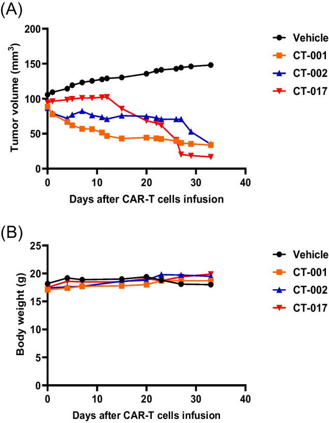
This study aimed to develop synthetic Claudin18.2 (CLDN18.2) chimeric antigen receptor (CAR)-T (CAR-T) cells as a treatment for advanced gastric cancer using lentiviral vector genetic engineering technology that targets the CLDN18.2 antigen and simultaneously overcomes the immunosuppressive environment caused by programmed cell death protein 1 (PD-1). Synthetic CAR T cells are a promising approach in cancer immunotherapy but face many challenges in solid tumors. One of the major problems is immunosuppression caused by PD-1. CLDN18.2, a gastric-specific membrane protein, is considered a potential therapeutic target for gastric and other cancers. In our study, CLDN18.2 CAR was a second-generation CAR with inducible T-cell costimulatory (CD278), and CLDN18.2-PD1/CD28 CAR was a third-generation CAR, wherein the synthetic PD1/CD28 chimeric-switch receptor (CSR) was added to the second-generation CAR. In vitro, we detected the secretion levels of different cytokines and the killing ability of CAR-T cells. We found that the secretion of cytokines such as interferon-gamma (IFN-γ) and tumor necrosis factor-alpha (TNF-α) secreted by three types of CAR-T cells was increased, and the killing ability against CLDN18.2-positive GC cells was enhanced. In vivo, we established a xenograft GC model and observed the antitumor effects and off-target toxicity of CAR-T cells. These results support that synthetic anti-CLDN18.2 CAR-T cells have antitumor effect and anti-CLDN18.2-PD1/CD28 CAR could provide a promising design strategy to improve the efficacy of CAR-T cells in advanced gastric cancer.

Keywords: CAR-T; Claudin18.2; Gastric cancer; Lentiviral vector; Synthetic PD1/CD28 receptor.


Conflict of interest statement

The authors have no financial conflicts of interest to declare.

Figures

Fig. 1
Fig. 1
The structure of synthetic CLDN18.2 CARs. A and B Schematic illustrations of CAR structures of CT-001, CT-017 (A), and CT-002 (B). CE Components of the synthetic CAR constructs CT-001, CT-017, and CT-002. Three individual CAR constructs include antigen-binding domain (anti-CLDN18.2 scFv), hinge domain (CD8α), transmembrane domain (ICOS), costimulatory domain (ICOS/CD3ζ), P2A, and chimeric-switch receptor (PD1/CD28) but have different composition and arrangement of transgenes and result in different sizes. The transgene sequences of the CT-001, CT-002, and CT-017 vectors are 2236, 2718, and 2352 bp, respectively. CT-001 and CT-017 have the same structure, except for the variation in the composition and arrangement of the PD1/CD28 CSR. CT-002 has an additional extracellular domain of the PD-1 receptor compared with CT-001. SP, signal peptide; TM, transmembrane domain; SD, stimulatory domain; P2A, 2A peptide; CSR, chimeric-switch receptor
Fig. 2
Fig. 2
The subcloning of plasmids, pCT-001, pCT-002, and pCT-017. The DNA sequences of the self-designed CAR transgene were synthesized and subcloned into a third-generation lentiviral vector modified from the pLenti-EF1a-Backbone (NG). A Lentiviral plasmid maps of pCT-001, pCT-002, and pCT-017. For pCT-001 and pCT-002, the synthesized DNA fragments were subcloned into a lentiviral vector using XbaI and XhoI, while the EZ-FusionTM HT Cloning method was used to generate pCT-017. The total sizes of pCT-001, pCT-002, and pCT-017 are 10169, 10664, and 10293 bp, respectively. B Electrophoresis of plasmids, pCT-001, pCT-002, and pCT-017. Plasmids were prepared from each clone and examined using DNA electrophoresis. The insertions and sequences of the synthesized DNA fragments were confirmed by sequencing
Fig. 3
Fig. 3
The characteristics of CLDN18.2 CAR-T cells. A Expression of CAR on T cells. Activated T cells were transduced using lentivirus encoding synthetic CLDN18.2-CAR constructs, and CAR expression level was assessed using flow cytometry after staining with FITC-labeled goat anti-mouse IgG F(ab’)2. B Proliferation curves of CAR-T cells and non-transduced T cells. In all cases, the cells expanded 200-fold 11 days post-transduction
Fig. 4
Fig. 4
Expression of Claudin18.2 in cancer cell lines. Claudin18.2 expression on cancer cells was examined by staining with Claudin18.2 recombinant human monoclonal antibody (Invitrogen) and goat anti-human IgG Fc secondary antibody-PE (Invitrogen). Claudin18.2 expression levels were 30.03% in NCI-N87, 38.63% in AGS, and 30.82% in KATO-III. High expression levels of Claudin18.2 are shown in the Claudin18.2 over-expressing cell lines NCI-N87-C18.2 (76.03%), AGS-C18.2 (99.68%), and KATO-III-C18.2 (99.73%)
Fig. 5
Fig. 5
Cytotoxicity and cytokine release of CLDN18.2 CAR-T cells in the presence of Claudin18.2-positive tumor cells. A Target cells were co-cultured with different effector cells at the indicated E:T ratios for 24 h. Cytotoxicity was quantified based on the LDH release. Data represent the mean ± SD (n = 3), and p-values were calculated using post-hoc tests after ANOVA. B CLDN18.2 CAR-T cells were co-cultured with the target cells for 24 h, and the supernatants were collected for ELISA. Data represent the mean ± SD (n = 3), and p-values were calculated using post-hoc tests after ANOVA. *p < 0.05, **p < 0.01, ***p < 0.001
Fig. 6
Fig. 6
Bioluminescence imaging of mice injected with CAR-T cells. A The mice were imaged using an IVIS Spectrum in vivo imaging system (PerkinElmer). Bioluminescence was visualized in vivo after intraperitoneal injection of D-luciferin (15 mg/ml, 200 μl/head) and analyzed using the image software (Living Image Version 4.7.2.20319) of the imaging system. G1, non-transduced T-cell administration group; G2, untreated group; G3, CT-017 CAR-T cell administration group; G4, CT-001 CAR-T cell administration group; G5, CT-002 CAR-T cell administration group. CAR-T cells were injected intratumorally (IT) into xenograft mice. Seven days after the first IT injection, they were injected intravenously (IV) through the tail vein. Bioluminescence from cancer cells significantly decreased in the CAR-T cell administration group. B Quantification of the total flux of light in the luminescence images. The results obtained using the IVIS were analyzed after setting the region of interest (ROI) and obtaining the total flux ([photons/seconds]). Each bar represents mean ± SD (n = 3). G1, non-transduced T-cell administration group; G2, untreated group; G3, CT-017 CAR-T cell administration group; G4, CT-001 CAR-T cell administration group; and G5, CT-002 CAR-T cells. The reduction of cancer cells in the G3, G4, and G5 CAR-T cell treatment groups was confirmed
Fig. 7
Fig. 7
The changes in tumor volume in mice after infusion of CAR-T cells. A Tumor volume. B Body weight. Vehicle: untreated group; CT-001, CT-001 CAR-T cell administration group; CT-002, CT-002 CAR-T cell administration group; CT-017, CT-017 CAR-T cell administration group. It was confirmed that cancer cells in the CAR-T cell administration groups were significantly reduced, which was consistent with the results of an anticancer efficacy test using BLI performed at 12, 20 and 33 days after the administration of CAR-T cells. Considering that there were no changes, such as weight loss, in any of the groups, it was determined that the anti-CLDN18.2 CAR-T cells used in this experiment had no in vivo toxicity

Similar articles

Cited by

References

    1. Abate-Daga, D., Rosenberg, S. A., & Morgan, R. A. (2014). Pancreatic cancer: Hurdles in the engineering of CAR-based immunotherapies. Oncoimmunology,3, e29194. 10.4161/onci.29194 - DOI - PMC - PubMed
    1. Alzubi, J., Dettmer-Monaco, V., Kuehle, J., Thorausch, N., Seidl, M., Taromi, S., Schamel, W., Zeiser, R., Abken, H., Cathomen, T., et al. (2020). PSMA-directed CAR T cells combined with low-dose docetaxel treatment induce tumor regression in a prostate cancer xenograft model. Molecular Therapy Oncolytics,18, 226–235. 10.1016/j.omto.2020.06.014 - DOI - PMC - PubMed
    1. Bates, P. D., Rakhmilevich, A. L., Cho, M. M., Bouchlaka, M. N., Rao, S. L., Hales, J. M., Orentas, R. J., Fry, T. J., Gilles, S. D., Sondel, P. M., et al. (2021). combining immunocytokine and ex vivo activated NK cells as a platform for enhancing graft-versus-tumor effects against GD2+ murine neuroblastoma. Frontiers in Immunology,12, 668307. 10.3389/fimmu.2021.668307 - DOI - PMC - PubMed
    1. Beckett, A. N., Chockley, P., Pruett-Miller, S. M., Nguyen, P., Vogel, P., Sheppard, H., Krenciute, G., Gottschalk, S., & DeRenzo, C. (2023). CD47 expression is critical for CAR T-cell survival in vivo. Journal for Immunotherapy of Cancer,11, e005857. 10.1136/jitc-2022-005857 - DOI - PMC - PubMed
    1. Bielamowicz, K., Fousek, K., Byrd, T. T., Samaha, H., Mukherjee, M., Aware, N., Wu, M. F., Orange, J. S., Sumazin, P., Man, T. K., et al. (2018). Trivalent CAR T cells overcome interpatient antigenic variability in glioblastoma. Neuro-Oncology,20, 506–518. 10.1093/neuonc/nox182 - DOI - PMC - PubMed

MeSH terms

LinkOut - more resources