Review
. 2025 Mar;28(2):506-518.
doi: 10.1111/vop.13219. Epub 2024 May 3.

Techniques for subretinal injections in animals

Affiliations
Review

Techniques for subretinal injections in animals

Ryan F Boyd et al. Vet Ophthalmol. 2025 Mar.

Abstract

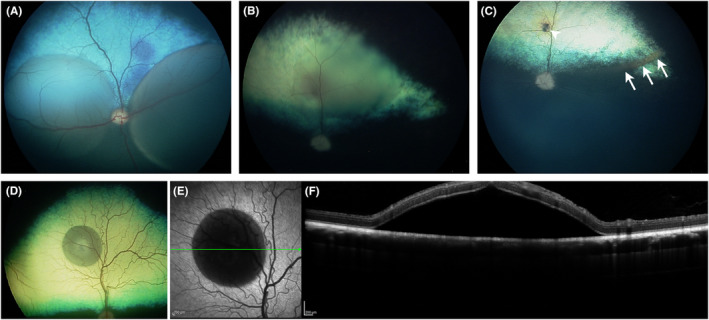
Subretinal injections are not commonly performed during clinical treatment of animals but are frequently used in laboratory animal models to assess therapeutic efficacy and safety of gene and cell therapy products. Veterinary ophthalmologists are often employed to perform the injections in the laboratory animal setting, due to knowledge of comparative ocular anatomy between species and familiarity with operating on non-human eyes. Understanding the different approaches used for subretinal injection in each species and potential complications that may be encountered is vital to achieving successful and reproducible results. This manuscript provides a summary of different approaches to subretinal injections in the most common animal model species, along with information from published literature and experience of the authors to educate novice or experienced surgeons tasked with performing these injections for the first time.

Keywords: animal model; retina; subretinal injection.


Conflict of interest statement

The authors declare no conflicts of interest.

Figures

FIGURE 1
FIGURE 1
Rodent intravitreal contact lens. A custom rodent intravitreal lens has been placed on the cornea using methylcellulose coupling gel, facilitating clear visualization of the retina and needle approach for subretinal injection in this albino rat.
FIGURE 2
FIGURE 2
Approaches for subretinal injection. Schematic of a rodent eye illustrating the needle path taken for transvitreal (A) and trans‐scleral (B) approaches. Note that a sharp beveled needle is required for a standard trans‐scleral approach to facilitate scleral and choroidal dissection, while a blunt needle may be used for the transvitreal approach following creation of a pilot sclerotomy hole.
FIGURE 3
FIGURE 3
Multiple subretinal blebs in a non‐human primate. The low lens:vitreous volume ratio in primates enables the surgeon to easily access all quadrants of the retina from a single approach. In this case, four 30 μL subretinal blebs were created to compare efficiency of four different viral vector solutions in a single eye, reducing the number of animals required for the investigation.
FIGURE 4
FIGURE 4
Subretinal injections in dogs. (A) Fundus image immediately after creating two subretinal blebs in a 3‐month‐old dog with normal retinal thickness. (B) Subretinal injection in a dog with moderate degree of retinal thinning due to PRA. The bleb is flatter than those created in the dog in (A). (C) Same dog as in (B) 24 h after injection. The bleb has flattened and there is some subretinal debris at the ventral edge of the bleb (indicated by arrows). The retinotomy site is indicated by the arrowhead. (D–F) Single small subretinal bleb in a normal dog. (D) Color RetCam image. (E) Confocal scanning laser ophthalmoscope (cSLO) infrared (IR) image. The green line indicates the plane of the spectral domain optical coherence tomography (SD‐OCT) cross‐sectional image shown in (F).
FIGURE 5
FIGURE 5
Subretinal injections in a cat. Cat was injected with an adeno‐associated viral vector delivering a green fluorescent protein marker gene. (A) Fundus prior to injection. (B) Image immediately post‐injection. (C–E) Fundus images 1‐, 2‐ and 3‐day post‐injection. These show that the subretinal fluid is cleared rapidly allowing retinal reattachment. The retinotomy site is indicated by the arrowhead in (E). Note also the subretinal debris accumulated at the ventral (dependent) portion of the bleb indicated by arrows in (E). (F) Fluorescent imaging showing GFP expression of the transduced retinal cells in the region of the bleb.
FIGURE 6
FIGURE 6
Retinal degeneration following subretinal injection in a rabbit. The thin retina of the rabbit is not conducive to recovery from routine subretinal injection, retinal degeneration is often observed. (A) Predose IR cSLO, (left panel) and SD‐OCT (right panel) images showing normal outer nuclear layer thickness in the rabbit (white arrow, hyporeflective layer). (B) Immediate post‐dose images following 70 μL subretinal injection, showing a subretinal bleb on cSLO with separation of the outer retina from the underlying RPE and choroid on SD‐OCT (white dashed lines). (C) Images taken 30 days post‐dose demonstrate diffuse outer nuclear layer thinning (white arrow) with central outer retinal folds (white arrowhead) within the subretinal dosing site.
FIGURE 7
FIGURE 7
Subretinal injection in a rabbit. If subretinal injection is performed in the rabbit, the subretinal cannula should be placed along the inferior margin of the myelinated medullary nerve fibers, which most often leads to a subretinal bleb that expands in an inferior direction to involve the visual streak, as shown in this photograph taken immediately following injection.
FIGURE 8
FIGURE 8
RPE detachment in the minipig. IR cSLO (left panel) and SD‐OCT (right panel) images taken 7 days following subretinal injection in a minipig eye. The small 10–20 μL subretinal bleb showed evidence of diffuse RPE detachment during bleb initiation, so the cannula was repositioned and a second subretinal bleb was created in a different quadrant. Note the accumulation of subretinal material within the central bleb that is hyporeflective on the cSLO image and hyper‐reflective on SD‐OCT (white arrowhead), likely representing accumulation of detached RPE. The surrounding bleb region is hyper‐reflective on cSLO, with thinning of the hyper‐reflective RPE layer on SD‐OCT (white arrows demonstrating bleb margins).

Similar articles

Cited by

References

    1. Haupert CL, McCuen BW, Jaffe GJ, et al. Pars plana vitrectomy, subretinal injection of tissue plasminogen activator, and fluid–gas exchange for displacement of thick submacular hemorrhage in age‐related macular degeneration. Am J Ophthalmol. 2001;131:208‐215. - PubMed
    1. Bainbridge JW, Smith AJ, Barker SS, et al. Effect of gene therapy on visual function in Leber's congenital amaurosis. N Engl J Med. 2008;358:2231‐2239. - PubMed
    1. Maguire AM, Simonelli F, Pierce EA, et al. Safety and efficacy of gene transfer for Leber's congenital amaurosis. N Engl J Med. 2008;358:2240‐2248. - PMC - PubMed
    1. Russell S, Bennett J, Wellman JA, et al. Efficacy and safety of voretigene neparvovec (AAV2‐hRPE65v2) in patients with RPE65‐mediated inherited retinal dystrophy: a randomised, controlled, open‐label, phase 3 trial. Lancet. 2017;390:849‐860. - PMC - PubMed
    1. Cheng S‐Y, Punzo C. Update on viral gene therapy clinical trials for retinal diseases. Hum Gene Ther. 2022;33:865‐878. - PMC - PubMed

LinkOut - more resources