. 2024 May 21;5(5):101522.
doi: 10.1016/j.xcrm.2024.101522. Epub 2024 May 3.

Microglial CMPK2 promotes neuroinflammation and brain injury after ischemic stroke

Affiliations

Microglial CMPK2 promotes neuroinflammation and brain injury after ischemic stroke

Xin Guan et al. Cell Rep Med. .

Abstract

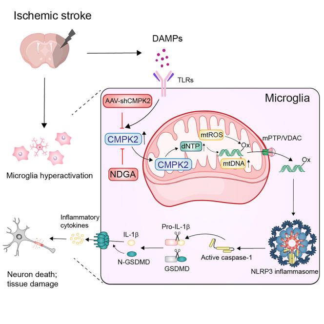
Neuroinflammation plays a significant role in ischemic injury, which can be promoted by oxidized mitochondrial DNA (Ox-mtDNA). Cytidine/uridine monophosphate kinase 2 (CMPK2) regulates mtDNA replication, but its role in neuroinflammation and ischemic injury remains unknown. Here, we report that CMPK2 expression is upregulated in monocytes/macrophages and microglia post-stroke in humans and mice, respectively. Microglia/macrophage CMPK2 knockdown using the Cre recombination-dependent adeno-associated virus suppresses the inflammatory responses in the brain, reduces infarcts, and improves neurological outcomes in ischemic CX3CR1Cre/ERT2 mice. Mechanistically, CMPK2 knockdown limits newly synthesized mtDNA and Ox-mtDNA formation and subsequently blocks NLRP3 inflammasome activation in microglia/macrophages. Nordihydroguaiaretic acid (NDGA), as a CMPK2 inhibitor, is discovered to reduce neuroinflammation and ischemic injury in mice and prevent the inflammatory responses in primary human monocytes from ischemic patients. Thus, these findings identify CMPK2 as a promising therapeutic target for ischemic stroke and other brain disorders associated with neuroinflammation.

Keywords: CMPK2; NLRP3 inflammasome; Ox-mtDNA; human PBMC cells; microglia; stroke.


Conflict of interest statement

Declaration of interests The authors declare no competing interests.

Figures

None
Graphical abstract
Figure 1
Figure 1
Peripheral blood expression of CMPK2 is elevated and positively correlative with infarct volume in human ischemic subjects (A) Quantification for CMPK2 gene expression (control = 21, stroke = 31). (B) Representative magnetic resonance imaging (MRI) images (diffusion-weighted imaging) of patients with stroke with CMPK2 gene expression (fold change < 10 and fold change > 10). (C) The correlation analysis was performed between CMPK2 transcript levels in blood and infarct volume in patients with stroke. n = 20. (D) The correlation analysis was performed between CMPK2 transcript levels in blood and NIHSS score in patients with stroke. n = 20. (E and F) Representative immunofluorescence images and quantification of 8-OHdG expression levels in the peripheral blood of ischemic patients. Scale bar: 20 μm. n = 6. (G and H) Representative immunofluorescence images and quantification of CMPK2 expression levels in the peripheral blood of patients with acute ischemic stroke. Scale bar: 20 μm. n = 6. Results are represented as means ± SD. ∗∗∗p < 0.001 vs. non-stroke control subjects.
Figure 2
Figure 2
CMPK2 expression is mainly upregulated in microglia/macrophages of the brain after ischemia onset (A and B) Western blotting images and quantification for CMPK2 protein expression in the peri-infarct region of sham-operated and ischemic mice at 3 h, 12 h, 1 day, 3 days, and 7 days following operation. n = 6. (C and D) Representative immunofluorescence images and quantitative analysis of CMPK2+ Iba1+ cells in the peri-infarct region of the ischemic mice. Scale bar: 50 μm. n = 6. (E and F) Western blotting images and quantification for CMPK2 protein expression in microglia/macrophages isolated from the peri-infarct region with CD11b+ magnetic beads at 24 h following operation. n = 6. (G and H) Western blotting images and quantification for CMPK2 expression in primary mouse microglia exposed to OGD conditions for 0, 2, 4, 6, 9, or 12 h. n = 4. (I and J) Western blotting images and quantification for CMPK2 expression in primary mouse microglia, astrocytes, or cortical neurons exposed to OGD conditions for 0 and 6 h. n = 6. Results are represented as means ± SD. ∗∗p < 0.01 and ∗∗∗p < 0.001 vs. sham or 0 h group.
Figure 3
Figure 3
Microglia/macrophage CMPK2 silencing reduces brain ischemic volume and improves short-term neurological function in the mouse tMCAO model (A) Schematic diagram about experimental strategy for CMPK2 knockdown. (B) Representative immunofluorescence images of mCherry+ cells stained with Iba1. Scale bar: 50 μm. n = 4. (C) Representative immunofluorescence images of mCherry+ cells stained with Iba1 and CMPK2. Scale bar: 20 μm. n = 6. (D) Western blotting images for CMPK2 protein expression in microglia/macrophages isolated from the brain of AAV-shCon and AAV-shCMPK2 CX3CR1Cre/ERT2 mice with CD11b+ magnetic beads. n = 6. (E and F) Regional cerebral blood flow. Results are expressed as means ± SD. n = 4. (G and H) AAV-shCMPK2 administration reduced cerebral infarct volume and promoted neurological functions in CX3CR1Cre/ERT2 male mice. n = 12. (I and J) AAV-shCMPK2 administration toned down cerebral infarct volume and improved neurological function in the CX3CR1Cre/ERT2 female mice. n = 8. Results are represented as means ± SD. ∗∗∗p < 0.001 vs. sham plus AAV-shCon group and ###p < 0.001 vs. tMCAO plus AAV-shCon group.
Figure 4
Figure 4
Microglia/macrophage CMPK2 silencing reduces brain infarction and improves long-term functional performances in the mouse tMCAO model (A) Schematic diagram about experimental procedures. (B and C) Representative T2-weighted MRI images of CX3CR1Cre/ERT2 mice on day 14 post-tMCAO. n = 4 or 5. (D) The long-term functional performances were investigated in CX3CR1Cre/ERT2 mice. n = 8. (E) The survival proportions. n = 12–15. Results are represented as means ± SD. ∗∗p < 0.01 and ∗∗∗p < 0.001 vs. sham plus AAV-shCon group and #p < 0.05, ##p < 0.01, and ###p < 0.001 vs. tMCAO plus AAV-shCon group.
Figure 5
Figure 5
Microglia/macrophage CMPK2 silencing promotes microglial ramification and disrupts Ox-mtDNA-induced NLRP3 inflammasome activation in the brain of ischemic mice (A) Imaris-based 3D reconstruction images of microglia immunofluorescently stained with Iba1. Scale bar: 15 μm. (B) The process length and the number of branches of microglia stained with Iba1 in AAV-shCon and AAV-shCMPK2 CX3CR1Cre/ERT2 mice at 3 days after ischemia. n = 10. Results are represented as means ± SD. (C–F) Representative immunofluorescence images and quantification of microglia/macrophages stained with 8-OHdG and quantitative analysis of 8-OHdG+ Iba1+ cells in the peri-infarct region of CX3CR1Cre/ERT2 mice at 24 and 72 h after ischemia. Scale bar: 50 μm. n = 6. (G) Western blotting images and quantification for cleaved caspase-1, mature IL-1β, and N-GSDMD in the peri-infarct region of AAV-shCon and AAV-shCMPK2 CX3CR1Cre/ERT2 mice at 3 days following ischemic stroke. n = 6. Results are represented as means ± SD. ∗∗∗p < 0.001 vs. sham plus AAV-shCon group and ###p < 0.001 vs. tMCAO plus AAV-shCon group.
Figure 6
Figure 6
The CMPK2-specific inhibitor NDGA suppresses Ox-mtDNA formation and NLRP3 inflammasome activation in microglia in vitro (A) The molecule structure of nordihydroguaiaretic acid (NDGA) and quantification of CMPK2 kinase assay with indicated concentrations of NDGA. n = 3. (B) The real-time binding kinetics of NDGA to immobilized CMPK2 protein were examined by SPR assay with the KD value of 0.6 μM. (C) NDGA reduced the protein expression of cleaved caspase-1, N-GSDMD, and mature IL-1β in the cell supernatant of LPS-primed microglia cells followed by ATP stimulation. (D and E) NDGA prevented LDH release in microglia subjected to LPS plus ATP stimulation or OGD model. n = 4. Results are represented as means ± SD. ∗∗∗p < 0.001 vs. control (CN) group and ##p < 0.01 and ###p < 0.001 vs. LPS plus ATP or OGD group. (F) NDGA decreased the Cox-1, CYTB, and D-loop expression in primary mouse microglia cells. n = 3. Results are represented as means ± SD. ∗∗∗p < 0.001 vs. CN group and ##p < 0.01 and ###p < 0.001 vs. LPS group. (G) NDGA reduced the newly synthesized DNA, as visualized by EdU labeling in primary mouse microglia cells. Scale bar: 5 μm. n = 6. (H) NDGA reduced 8-OHdG expression in LPS-primed microglia followed by ATP stimulation. Scale bar: 20 μm. n = 4.
Figure 7
Figure 7
The CMPK2 inhibitor NDGA reduces brain ischemic volume and improves neurological performances in the mouse tMCAO model (A–C) NDGA reduced cerebral infarct volume and promoted short-term neurological functions in the male mice. n = 8. (D) NDGA improved long-term functional performances, including mNSS test, wire-hanging test, forelimb foot fault test, and rotarod test in the male mice. n = 8. (E) NDGA improved long-term survival proportions in the male mice. n = 12–15. Results are represented as means ± SD. ∗p < 0.05, ∗∗p < 0.01, and ∗∗∗p < 0.001 vs. sham group and #p < 0.05, ##p < 0.01, and ###p < 0.001 vs. tMCAO plus vehicle group.

References

    1. Owolabi M.O., Thrift A.G., Mahal A., Ishida M., Martins S., Johnson W.D., Pandian J., Abd-Allah F., Yaria J., Phan H.T., et al. Primary stroke prevention worldwide: translating evidence into action. Lancet Public Health. 2022;7:e74–e85. doi: 10.1016/S2468-2667(21)00230-9. - DOI - PMC - PubMed
    1. Yu H., Chen X., Guo X., Chen D., Jiang L., Qi Y., Shao J., Tao L., Hang J., Lu G., et al. The clinical value of serum xanthine oxidase levels in patients with acute ischemic stroke. Redox Biol. 2023;60 doi: 10.1016/j.redox.2023.102623. - DOI - PMC - PubMed
    1. Barclay R.E., Stevenson T.J., Poluha W., Semenko B., Schubert J. Mental practice for treating upper extremity deficits in individuals with hemiparesis after stroke. Cochrane Database Syst. Rev. 2020;5 doi: 10.1002/14651858.CD005950.pub5. - DOI - PMC - PubMed
    1. Global, regional, and national burden of stroke and its risk factors, 1990-2019: a systematic analysis for the Global Burden of Disease Study 2019. Lancet Neurol. 2021;20:795–820. doi: 10.1016/S1474-4422(21)00252-0. - DOI - PMC - PubMed
    1. Wang R., Zhu Y., Liu Z., Chang L., Bai X., Kang L., Cao Y., Yang X., Yu H., Shi M.-J., et al. Neutrophil extracellular traps promote tPA-induced brain hemorrhage via cGAS in mice with stroke. Blood. 2021;138:91–103. doi: 10.1182/blood.2020008913. - DOI - PMC - PubMed

MeSH terms